38 head and neck muscle diagram
Blank Head and Neck Muscles Diagram. Find this Pin and more on Summer by Rainey Stoner. Head Muscles. Facial Muscles. Nursing Notes. Nursing Tips. Neck Muscle Anatomy. Muscle Diagram. Anatomy Bones. Human muscle system, the muscles of the human body that work the skeletal system, that are under voluntary control, and that are concerned with movement, posture, and balance. Broadly considered, human muscle—like the muscles of all vertebrates—is often divided into striated muscle, smooth muscle, and cardiac muscle.
Head and neck muscles labeled anatomical diagram, facial vector illustration with female face, health care educational information poster. Fitness and beauty related. anatomy of neck and shoulder stock illustrations

Head and neck muscle diagram
Dec 14, 2021 · Muscles of the neck (Musculi cervicales) The muscles of the neck are muscles that cover ... Definition And Function. A Large Group Of Muscles In Th ... Anterior muscles of the neck. Superficial muscles: Platysma, ... Lateral (vertebral) muscles of ... Scalene muscles: Anterior scal ... Sep 30, 2021 · Head and neck (anterior view) The head and neck are two examples of the perfect anatomical marriage between form and function, mixed with a dash of complexity. The neck is resilient enough to sustain a five kilogram weight 24/7, yet sufficiently mobile to move it in several directions. Start studying Head and Neck Muscles. Learn vocabulary, terms, and more with flashcards, games, and other study tools.
Head and neck muscle diagram. Skin. The head and neck is covered in skin and its appendages, termed the integumentary system.These include hair, sweat glands, sebaceous glands, and sensory nerves.The skin is made up of three microscopic layers: epidermis, dermis, and hypodermis.The epidermis is composed of stratified squamous epithelium and is divided into the following five sublayers or strata, listed in order from outer ... Lateral View - Head, Neck, and Shoulder Muscles . Lateral View - Pectoral Girdle The cleidocervicalis is labeled clavotrapezius in your book. This figure illustrates the position of the transversus abdominus in relation to the internal and external oblique muscles. Skull (lateral view) Blank Diagram. Complete Diagram. Muscles of the Head and Neck. Cervical Plexus. Blank Diagram. Complete Diagram. Oral Cavity. Central Nervous System. Here are a number of highest rated Head And Neck Muscle Labeling pictures upon internet. We identified it from well-behaved source. Its submitted by dealing out in the best field. We acknowledge this kind of Head And Neck Muscle Labeling graphic could possibly be the most trending topic in the same way as we share it in google plus or facebook.
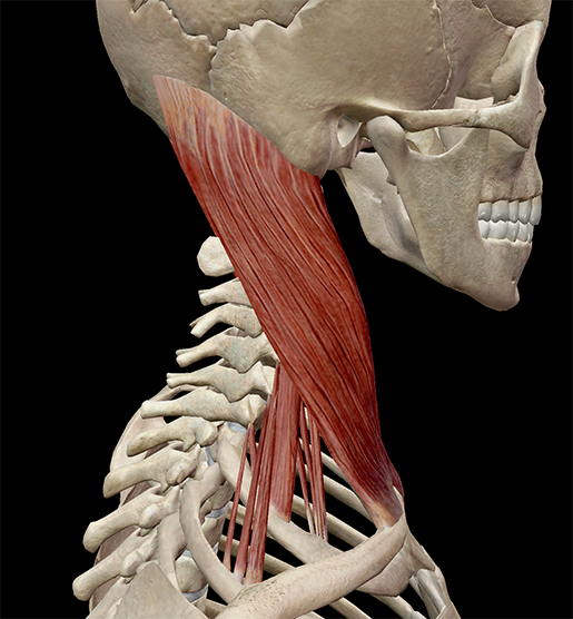
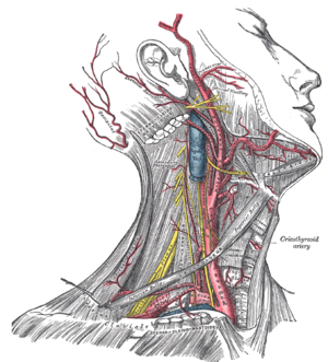
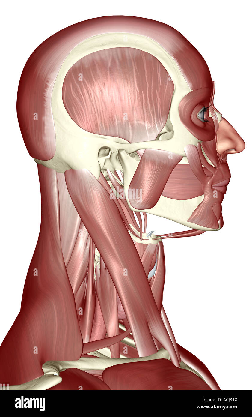
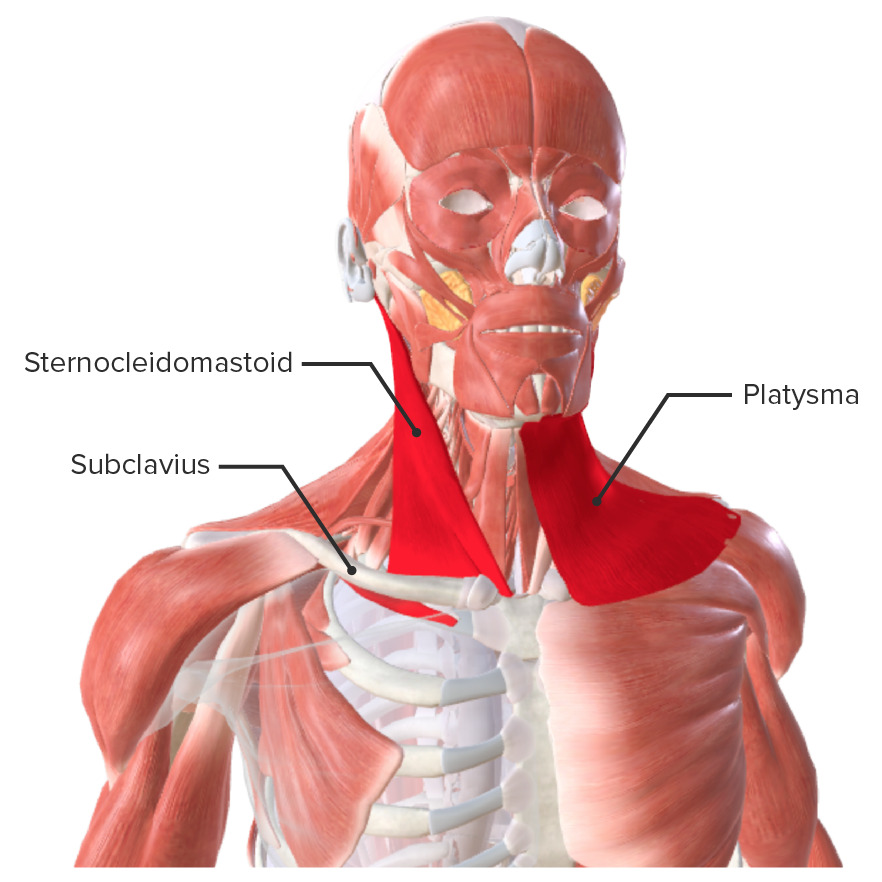
sternocleidomastoid. two-headed muscle located deep to platysma on anterolateral surface of neck, fleshy parts on either side of neck delineate limits of triangles, origin- manubrium and medial portion of clavicle, insertion- mastoid process and superior nuchal line of occipital bone, flexes and laterally rotates the head. scalene. Start studying Lateral Head and Neck (deeper muscles). Learn vocabulary, terms, and more with flashcards, games, and other study tools. Jan 21, 2018 · The neck is the start of the spinal column and spinal cord. The spinal column contains about two dozen inter-connected, oddly shaped, bony segments, called vertebrae. The neck contains seven of ... About this Quiz. This is an online quiz called Muscles of the Face, Head, and Neck. There is a printable worksheet available for download here so you can take the quiz with pen and paper.
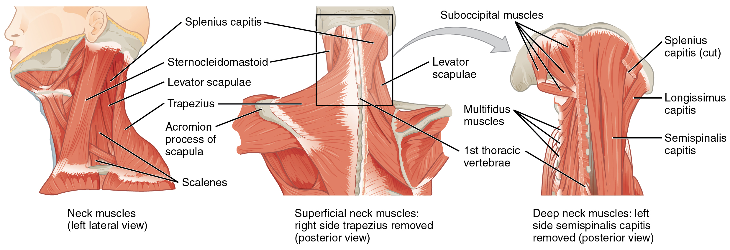
Feb 17, 2015 · Superficial muscles are the muscles closest to the skin surface and can usually be seen while a body is performing actions. Many in the neck help to stabilize or move the head. Some also create ... This Osmosis High-Yield Note provides an overview of Head and Neck Structure essentials. All Osmosis Notes are clearly laid-out and contain striking images, tables, and diagrams to help visual learners understand complex topics quickly and efficiently. Find more information about Head and Neck Structure by visiting the associated Learn Page. Head and neck muscles labeled anatomical diagram, facial vector illustration with female face, health care educational information. Illustration about movement, adult - 118338016 Instant anatomy is a specialised web site for you to learn all about human anatomy of the body with diagrams, podcasts and revision questions
11.4 Identify the skeletal muscles and give their origins ...
Cross-sectional labeled anatomy of the head and neck of the domestic cat on CT imaging (bones of the skull, cervical spine, mandible, hyoid bone, muscles of the neck, nasal cavity and paranasal sinuses, oral cavity, larynx)
Learn Muscle Anatomy: Scalene Muscles and Other Neck Anatomy
Feb 09, 2017 · Important exam questions on anatomy of head and neck- enumerate, draw labelled diagrams, short notes, long questions and applied anatomy questions.

Homo sapiens Neck Thorax Muscle Organ, others, hand, human ...
Muscles of Head and Neck. Anterior and Lateral Neck Muscles Blank Diagram Complete Diagram Supra/Infrahyoid Muscles Blank Diagram Complete Diagram Pharyngeal Constrictor Muscles Blank Diagram Complete Diagram Muscles of Facial Expression Blank Diagram Complete Diagram Muscles of Mastication Lateral Skull (Blank Diagram) Mastication 1(Complete ...

Amazon.com: Head and Neck Anatomical Chart: 9781587791482 ...
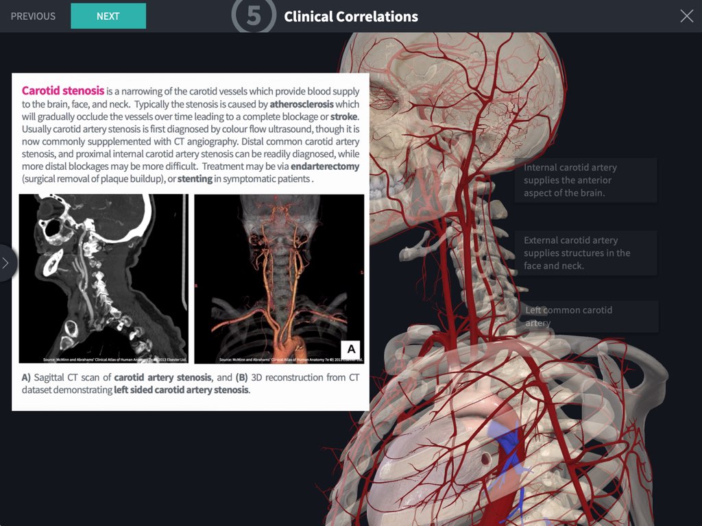
The neck is the bridge between the head and the rest of the body. It is located in between the mandible and the clavicle, connecting the head directly to the torso, and contains numerous vital structures. It contains some of the most complex and intricate anatomy in the body and is comprised of numerous organs and tissues with essential structure and function for normal physiology.

Head and Neck | Radiology Key
Blank Head and Neck Muscles Diagram. Why is it important to learn muscle anatomy? Muscle and anatomy are two words that are often heard when you are studying science. The human body consists of many muscles. If someone wants a healthy and good life, one must understand his body. How do you take care of a body if you don't know the anatomy?
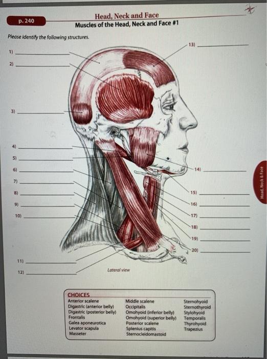
Solved p. 240 Head, Neck and Face Muscles of the Head, Neck ...
Jun 11, 2021 · The salivary glands are exocrine glands that make, modify and secrete saliva into the oral cavity. They are divided into two main types: the major salivary glands, which include the parotid, submandibular and sublingual glands, and the minor salivary glands, which line the mucosa of the upper aerodigestive tract and the overwhelming entirety of the mouth [1].

Primary Neck Cancers ‣ Anatomy
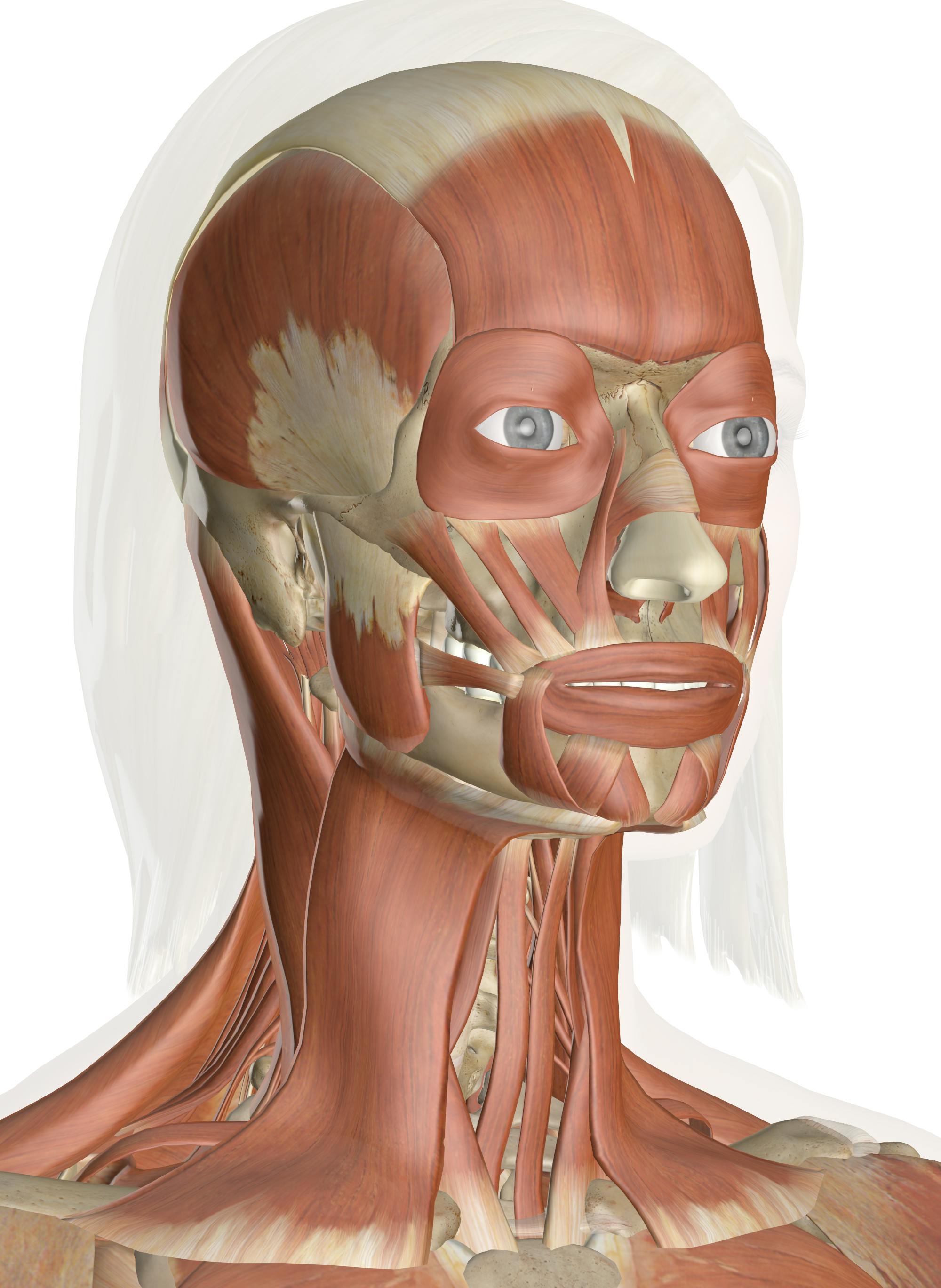
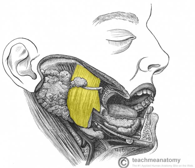
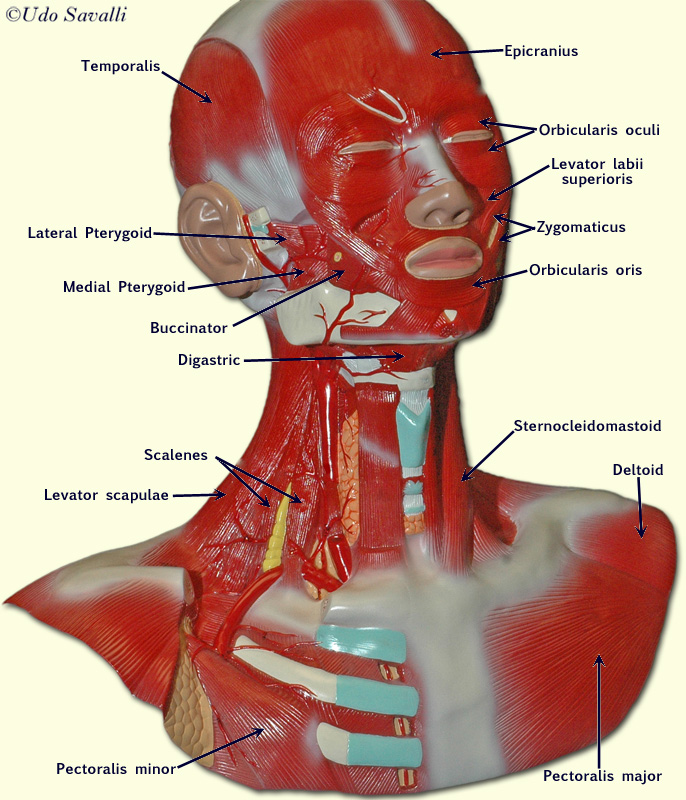
The primary muscles of mastication (chewing food) are the temporalis, medial pterygoid, lateral pterygoid, and masseter muscles. The four main muscles of mastication attach to the rami of the mandible and function to move the jaw (mandible). The cardinal mandibular movements of mastication are elevation, depression, protrusion, retraction, and side to side movement.
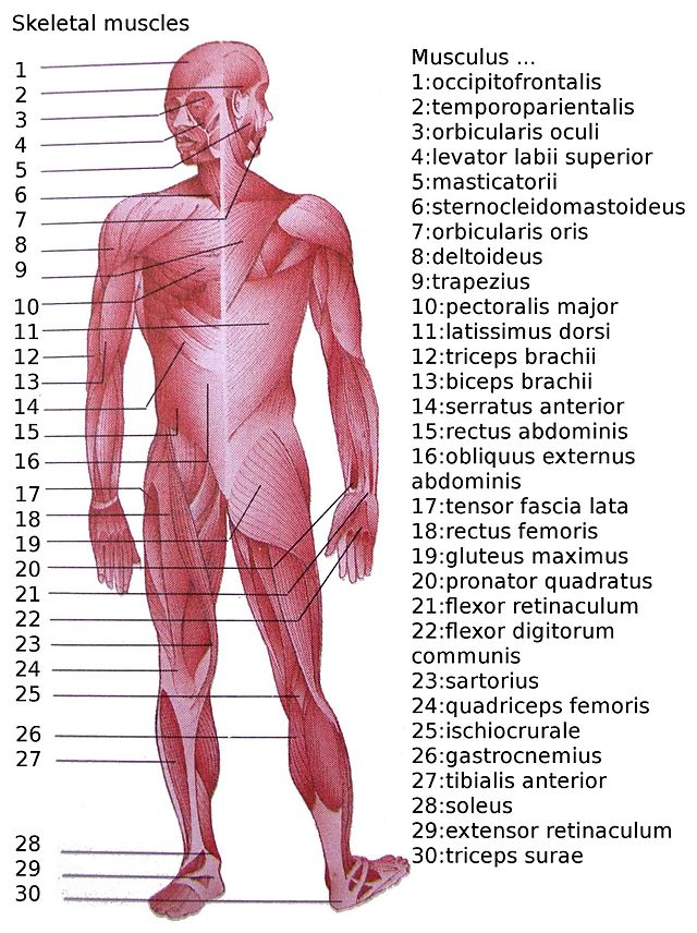
List of skeletal muscles of the human body - Wikipedia
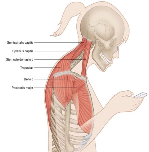
The neck muscles, including the sternocleidomastoid and the trapezius, are responsible for the gross motor movement in the muscular system of the head and neck. They move the head in every direction, pulling the skull and jaw towards the shoulders, spine, and scapula. Working in pairs on the left and right sides of the body, these muscles ...
Muscles of the Head and Neck - Anatomy Pictures and Information
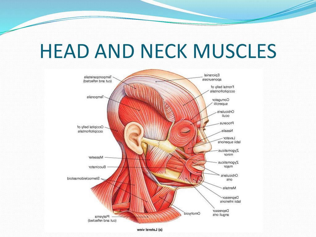
Muscles of the Head and Neck. Humans have well-developed muscles in the face that permit a large variety of facial expressions. Because the muscles are used to show surprise, disgust, anger, fear, and other emotions, they are an important means of nonverbal communication. Muscles of facial expression include frontalis, orbicularis oris, laris ...
Head and neck anatomy - Wikipedia
Muscles of the neck (Musculi cervicales) The muscles of the neck are muscles that cover the area of the neck.These muscles are mainly responsible for the movement of the head in all directions. They consist of 3 main groups of muscles: anterior, lateral and posterior groups, based on their position in the neck.The musculature of the neck is further divided into more specific groups based on a ...

627 Head And Neck Diagram Stock Photos, Pictures & Royalty ...
Download this Head And Neck Muscles Labeled Anatomical Diagram Facial Vector Illustration With Female Face Health Care Educational Information Poster vector illustration now. And search more of iStock's library of royalty-free vector art that features Muscle graphics available for quick and easy download.
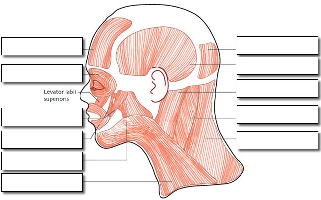
Label the Muscles of the Head
Jul 31, 2021 · The eyes are a set of sensory organs that play a crucial role in the visual system. The eyes are responsible for detecting light that enters the eyes. Then, the light gets converted into an image in the brain. The sensory and motor innervation of the eyes originate from six paired cranial nerves. These nerves work in sync to manifest movements, reflexes, and vision.

Anatomy of short neck muscles. Short Neck Muscles with Marked ...
This is an online quiz called Muscles of the Head, Neck, & Face. There is a printable worksheet available for download here so you can take the quiz with pen and paper. Your Skills & Rank. Total Points. 0. Get started! Today's Rank--0. Today 's Points. One of us! Game Points. 18.

Anatomy of the Head and Neck - Medical Illustrations showing ...
Neck muscles are bodies of tissue that produce motion in the neck when stimulated. The muscles of the neck run from the base of the skull to the upper back and work together to bend the head and ...

Head and Neck Cancer | NEJM
Start studying Head and Neck Muscles. Learn vocabulary, terms, and more with flashcards, games, and other study tools.

Gross Anatomy Of Skeletal Muscle The Muscular System Micro ...
Sep 30, 2021 · Head and neck (anterior view) The head and neck are two examples of the perfect anatomical marriage between form and function, mixed with a dash of complexity. The neck is resilient enough to sustain a five kilogram weight 24/7, yet sufficiently mobile to move it in several directions.
Gross Head and Neck Anatomy
Dec 14, 2021 · Muscles of the neck (Musculi cervicales) The muscles of the neck are muscles that cover ... Definition And Function. A Large Group Of Muscles In Th ... Anterior muscles of the neck. Superficial muscles: Platysma, ... Lateral (vertebral) muscles of ... Scalene muscles: Anterior scal ...
11.3 Axial Muscles of the Head, Neck, and Back – Douglas ...

Neck Muscles and Other Soft Tissues
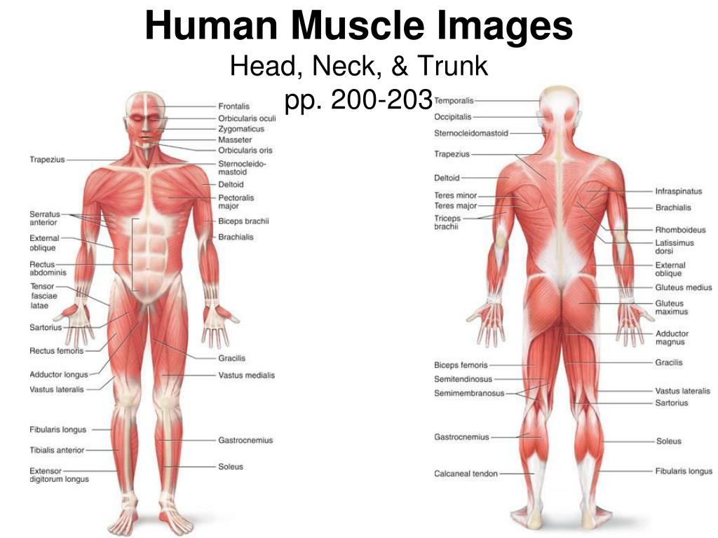
PPT - Human Muscle Images Head, Neck, & Trunk pp. 200-203 ...
Muscles of the Head - TeachMeAnatomy

Head and neck muscle anatomy
head | Definition & Anatomy | Britannica
Neck muscles anatomy: List, origins, insertions, action | Kenhub

BIO201-Head Neck Muscles
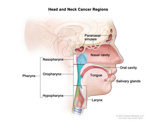
Head and Neck Cancers - National Cancer Institute
The muscles of the head neck and face Stock Photo - Alamy

Muscles of the Head & Neck | Anatomy Model - YouTube
6 Exercises to Reduce Stress and Strain in the Neck - IMPACT ...

Posterior triangle of the neck Head and neck anatomy ...
THORACIC AND HEAD/NECK MUSCLES - ppt download

Axial Muscles of the Head, Neck & Back: Structure, Movement ...
Muscles of the Neck | Concise Medical Knowledge

The muscles of the head and neck | Human Anatomy and ...

The head and neck muscles stock illustration. Illustration of ...
BIO201-Head Neck Muscles

Head and Neck Human Anatomy (Muscles)

Overview of the head and neck region - Knowledge @ AMBOSS
0 Response to "38 head and neck muscle diagram"
Post a Comment