41 muscle cell diagram labeled
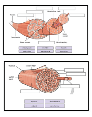
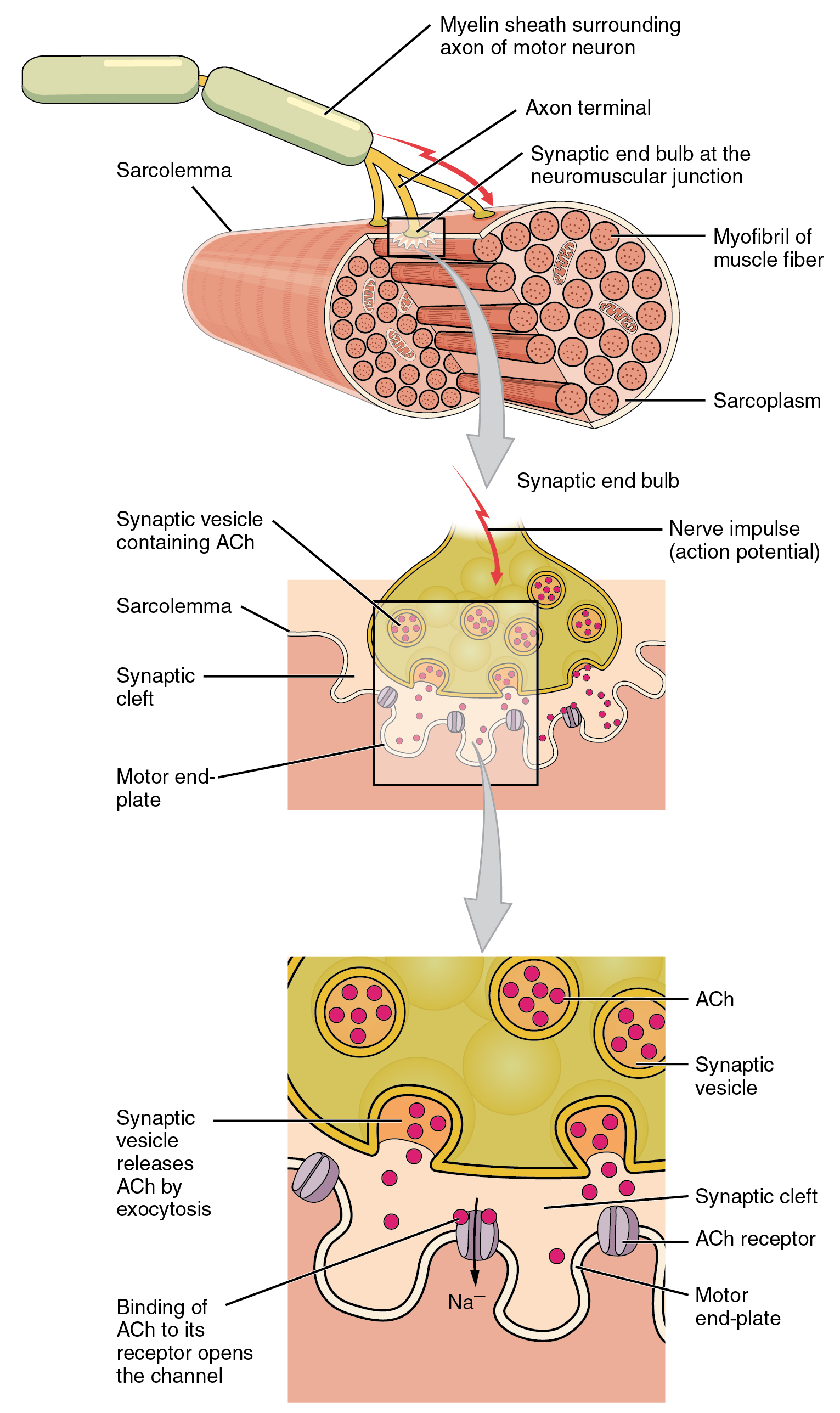
Muscle tissue - Wikipedia Skeletal muscle is broadly classified into two fiber types: Type I slow-twitch, and Type II fast-twitch muscle. Type I, slow-twitch, slow oxidative, or red muscle is dense with capillaries and is rich in mitochondria and myoglobin, giving the muscle tissue its characteristic red color. 10.2 Skeletal Muscle - Anatomy & Physiology Figure 10.2.2 - Muscle Fiber: A skeletal muscle fiber is surrounded by a plasma membrane called the sarcolemma, which contains sarcoplasm, the cytoplasm of muscle cells. A muscle fiber is composed of many myofibrils, which contain sarcomeres with light and dark regions that give the cell its striated appearance. The Sarcomere
Muscle Cell Structure - Biology MUSCLE CELL STRUCTURE - BIOLOGY. Each muscle fibre is covered by sarcolemma. Tunnel-like extensions pass through the muscle fibre from one side of it to the other in transverse sections through the diameter of the fibre. These tunnel-like extensions are known as transverse tubules. The nuclei of muscle cells are located at the edges of the ...
Muscle cell diagram labeled
Ultrastructure of Muscle - Skeletal - Sliding Filament ... Composition of Skeletal Muscle. A muscle cell is very specialised for its purpose. A single cell forms one muscle fibre, and its cell surface membrane is known as the sarcolemma.. T tubules are unique to muscle cells. These are invaginations of the sarcolemma that conduct charge when the cell is depolarised. Foot Diagram: Labeled Anatomy - Science Trends Jul 05, 2018 · The foot diagram has a complex structure made up of bones, ligaments, muscles, and tendons. Understanding the structure of the foot is best done by looking at a foot diagram where the anatomy has been labeled. If you would like to learn all the parts of the foot structure, you have come to the right place. 20 Unlabeled Muscle Diagram Worksheet | Worksheet From Home 20 Unlabeled Muscle Diagram Worksheet Label Muscles Worksheet unlabeled male reproductive system, unlabeled muscle fiber, unlabeled muscles, unlabeled muscular system image, unlabeled male reproductive system diagram, via: pinterest.com Numbering Worksheets for Kids. Kids are usually introduced to this topic matter during their math education.
Muscle cell diagram labeled. Skeletal Muscle Cell Quiz - PurposeGames.com This is an online quiz called Skeletal Muscle Cell There is a printable worksheet available for download here so you can take the quiz with pen and paper. From the quiz author Labels/Diagram based on Figure 6.3 "Anatomy of Skeletal Muscle Fiber" Your Skills & Rank Total Points 0 Get started! Today's Rank -- 0 Today 's Points One of us! Game Points Chapter 10 - Muscle Cell Labeling Flashcards - Quizlet Start studying Chapter 10 - Muscle Cell Labeling. Learn vocabulary, terms, and more with flashcards, games, and other study tools. Skeletal Muscle Histology Slide Identification and Labeled ... The skeletal muscle fibers are elongated, cylindrical and multinucleated cells whose length may vary in different animals. In this short guide, you will get a basic concept of skeletal muscle histology from the real slide and labeled diagram. You will also get the identification points of skeletal muscle histology slide with a little description here in this guide. Solved Muscle Using a labeled diagram, detail all the ... Anatomy and Physiology questions and answers Muscle Using a labeled diagram, detail all the events that occur in a muscle contraction/NMJ staring with an action potential reaches the axonal terminal of a motor neuron and ending with myosin disengages from actin in the muscle cell and Ach is broken down
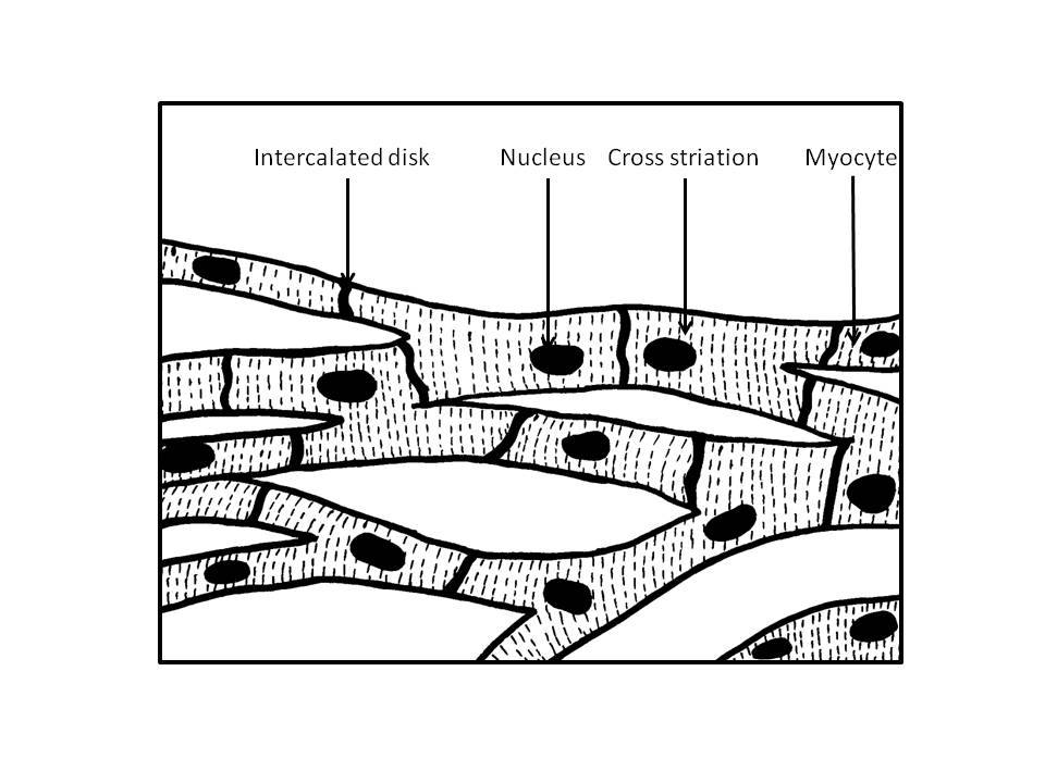
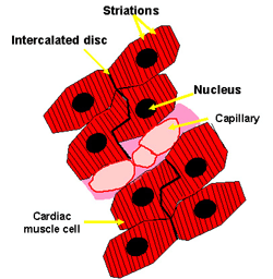
Metabolism of Carbohydrates: 10 Cycles (With Diagram) ADVERTISEMENTS: This article throws light upon the ten major pathways/cycles of carbohydrate metabolism. The ten pathways/cycles of carbohydrate metabolism are: (1) Glycolysis (2) Conversion of Pyruvate to Acetyl COA (3) Citric Acid Cycle (4) Gluconeogenesis (5) Glycogen Metabolism (6) Glycogenesis (7) Glycogenolysis (8) Hexose Monophosphate Shunt (9) Glyoxylate Cycle and (10) Photosynthesis ... 19.2 Cardiac Muscle and Electrical Activity - Anatomy and ... Figure 19.17 Cardiac Muscle (a) Cardiac muscle cells have myofibrils composed of myofilaments arranged in sarcomeres, T tubules to transmit the impulse from the sarcolemma to the interior of the cell, numerous mitochondria for energy, and intercalated discs that are found at the junction of different cardiac muscle cells. (b) A photomicrograph of cardiac muscle cells shows the nuclei and ... Muscle Cell (Myocyte): Definition, Function & Structure ... A muscle cell, known technically as a myocyte, is a specialized animal cell which can shorten its length using a series of motor proteins specially arranged within the cell. While several associated proteins help, actin and myosin form thick and thin filaments which slide past each other to contract small units of a muscle cell. Muscles Body Diagram Stock Illustrations - 564 Muscles ... Medical educational diagram and scheme with satellite cell and fusion of cells. Vector illustration about how. Head and neck muscles labeled anatomical diagram, facial vector illustration with female face, health care educational information. Poster. Fitness and beauty.
Skeletal Muscle - Anatomy & Physiology Skeletal muscle fibers can be quite large for human cells, with diameters up to 100 μ m and lengths up to 30 cm (11.8 in) in the Sartorius of the upper leg. During early development, embryonic myoblasts, each with its own nucleus, fuse with up to hundreds of other myoblasts to form the multinucleated skeletal muscle fibers. Muscular System Labeling Worksheet Muscular System Labeling Worksheet Somatic signals are sent from the cerebral cortex to nerves associated with specific skeletal muscles. This results in muscular hypertrophy which is a result in a increase of myofibrils as a result of increased exercise level of physical activity determined by the frequency of recruit ment and the load. Muscle cell - Wikipedia Structure. The unusual microscopic anatomy of a muscle cell gave rise to its own terminology. The cytoplasm in a muscle cell is termed the sarcoplasm; the smooth endoplasmic reticulum of a muscle cell is termed the sarcoplasmic reticulum; and the cell membrane in a muscle cell is termed the sarcolemma. The sarcolemma receives and conducts stimuli. Skeletal muscle cells Labelled diagram of a muscle cell | Human anatomy model ... Myofibril : Anatomy of Muscle Structure Myofibrils are cylindrical structures that extend along the complete length of each muscle cell. Each myofibril consists of two types of protein filaments - thin filaments and thick filaments. S shanon hernandez 86 followers More information Labelled diagram of a muscle cell
Labeled Sarcomere Diagram A sarcomere is the basic unit of striated muscle tissue. It is the repeating unit between two Z lines. Skeletal muscles are composed of tubular muscle cells which. Sarcomeres are composed of thick filaments and thin filaments. The thin filaments Look at the diagram above and realize what happens as a muscle contracts.
Primary Cell Culture: 3 Techniques (With Diagram) ADVERTISEMENTS: This article throws light upon the three types of technique used for primary cell culture. The three types of technique are: (1) Mechanical Disaggregation (2) Enzymatic Disaggregation and (3) Primary Explant Technique. Primary culture broadly involves the culturing techniques carried following the isolation of the cells, but before the first subculture. Primary cultures are […]
Muscle Anatomy Quiz - Registered Nurse RN Muscle anatomy quiz for anatomy and physiology! When you are taking anatomy and physiology you will be required to identify major muscles in the human body. This quiz requires labeling, so it will test your knowledge on how to identify these muscles (latissimus dorsi, trapezius, deltoid, biceps brachii, triceps brachii, brachioradialis, pectoralis major, serratus anterior, rectus abdominis, etc.).
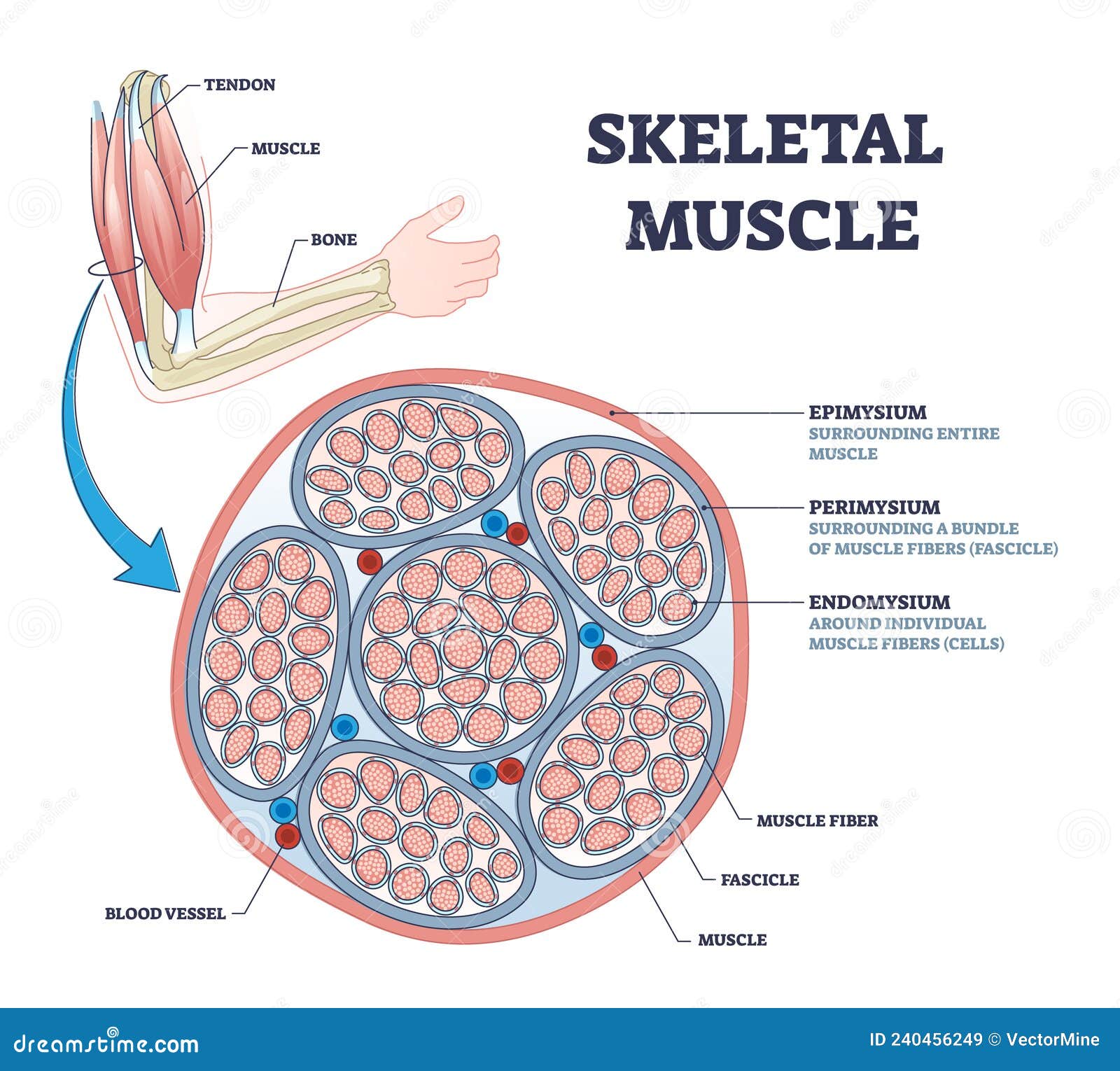
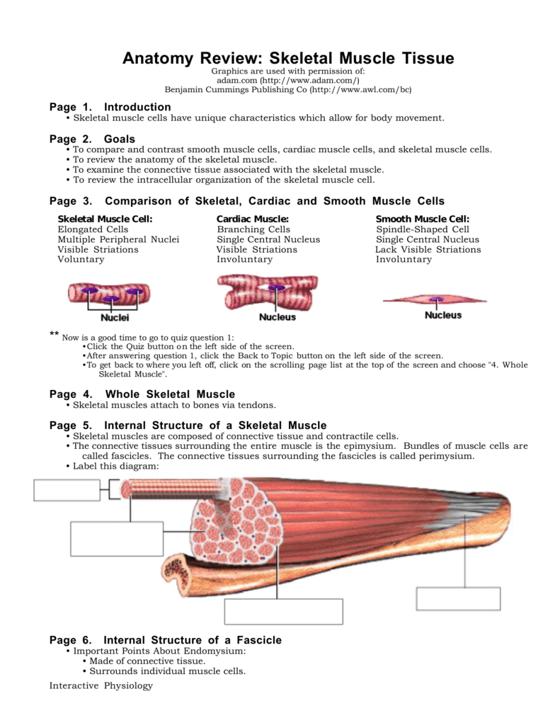
Muscle Cell | Definition, Anatomy, Types & Functions Muscles are composed of connective tissue and contractile cells. The connective tissues surrounding the entire muscle is the epimysium. Bundles of muscle cells are called fascicles. The connective tissues surrounding the fascicles are called perimysium. The fascicle is made of connective tissue which surrounds individual muscle cells.
PDF Anatomy Review: Skeletal Muscle Tissue • Skeletal muscle cells have unique characteristics which allow for body movement. Page 2. Goals • To compare and contrast smooth muscle cells, cardiac muscle cells, and skeletal muscle cells. • To review the anatomy of the skeletal muscle. • To examine the connective tissue associated with the skeletal muscle.
Structure and Function of the Skeletal Muscle Extracellular ... Sep 01, 2011 · Furthermore, multiple reaction monitoring may be used in the future to quantify proteins from labeled cell types in muscle. 107 This method can detect and quantify peptides in multiplex format using a triple quadrupole instrument. An advantage to this method is that it might be used to identify protein signals in fibrotic muscle that cause ...
Cardiomyocytes (Cardiac Muscle Cells)- Structure, Function ... · Label the cells for 30 minutes in the dark at room temperature using: Alexa Fluor 568 phalloidin - to stain actin SYTO 11 Green-Fluorescent Nucleic Acid - to stain the nucleus · Wash the cells using PBS three times (three minutes for each wash) · Maintain the cells in PBS containing antibiotics
Sarcomere Diagram Labeled Sarcomere The figure depicts the structure of a Sarcomere. (Each zone is labeled). They first. Start studying UNIT 5: Label the parts of the Sarcomere. Learn vocabulary, terms, and more with flashcards, games, and other study tools.A sarcomere is the basic functional within muscle cells. This unit is distinctive in some types of muscle tissue.
Printable Human Body Diagram - Studying Diagrams Aug 26, 2021 · The immune integumentary skeletal muscle and reproductive systems are also part of the human body. Human Body Systems Project Lift-the-Flap Model w Human Body Reading Passages Human Body Activities for Kids Everything included. The human body is an amazing and complex thing. Free Printable Human Body Diagram for Kids Labeled and Unlabeled.
Label the muscle cell diagram Quiz - PurposeGames.com This is an online quiz called Label the muscle cell diagram There is a printable worksheet available for download here so you can take the quiz with pen and paper. Your Skills & Rank Total Points 0 Get started! Today's Rank -- 0 Today 's Points One of us! Game Points 16 You need to get 100% to score the 16 points available Actions
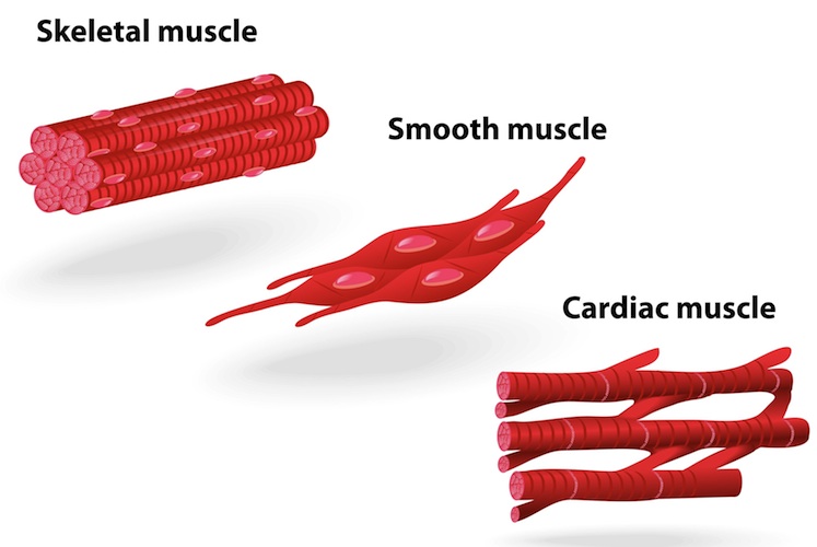
Types of muscle cells: Characteristics, location, roles ... There are 3 types of muscle cells in the human body; cardiac, skeletal, and smooth. Cardiac and skeletal myocytes are sometimes referred to as muscle fibers due to their long and fibrous shape. Cardiac muscle cells, or cardiomyocytes, are the muscle fibers comprise the myocardium, the middle muscular layer, of the heart.
Muscles Notes: Diagrams & Illustrations | Osmosis NOTES NOTES MUSCLES MUSCULAR SYSTEM ANATOMY & PHYSIOLOGY osms.it/muscle-anatomy-physiology Three types of muscle cell/tissue Skeletal, cardiac, smooth Differ in location, innervation, cell structure All cells excitable, extensible, elastic SKELETAL MUSCLE Attaches to bone/skin; mostly voluntary; maintains posture, stabilizes joints, generates heat Most muscles consist of belly (contracts ...
Learn all muscles with quizzes and labeled diagrams | Kenhub Labeled diagram View the muscles of the upper and lower extremity in the diagrams below. Use the location, shape and surrounding structures to help you memorize each muscle. Once you're feeling confident, it's time to test yourself. Unlabeled diagram See if you can label the muscles yourself on the worksheet available for download below.
PDF Muscle Cell Anatomy & Function Human Anatomy & Physiology: Muscle Physiology; Ziser Lecture Notes, 2006 1 Muscle Cell Anatomy & Function (mainly striated muscle tissue) General Structure of Muscle Cells (skeletal) several nuclei (skeletal muscle) skeletal muscles are formed when embryonic cells fuse together some of these embryonic cells remain in the adult and can replace
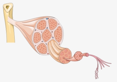
Structure of Skeletal Muscle | SEER Training Within the fasciculus, each individual muscle cell, called a muscle fiber, is surrounded by connective tissue called the endomysium. Skeletal muscle cells (fibers), like other body cells, are soft and fragile. The connective tissue covering furnish support and protection for the delicate cells and allow them to withstand the forces of contraction.
Label structure of skeletal muscle Diagram | Quizlet a long, filamentous organelle found within muscle cells that has a banded appearance tendon cordlike extension of connective tissue beyond the muscle, serving to attach it to the bone sarcolemma plasma membrane of a muscle cell epimysium just deep to the deep fascia (surrounds entire muscle) perimysium
Ileocecal valve - Wikipedia Microanatomy. The histology of the ileocecal valve shows an abrupt change from a villous mucosa pattern of the ileum to a more colonic mucosa. A thickening of the muscularis mucosa, [citation needed] which is the smooth muscle tissue found beneath the mucosal layer of the digestive tract.
Anatomy, Skeletal Muscle - StatPearls - NCBI Bookshelf The three main types of muscle tissue are skeletal, cardiac, and smooth muscle groups. [1][2][3]Skeletal muscle attaches to the bone by tendons, and together they produce all the movements of the body. The skeletal muscle fibers are crossed with a regular pattern of fine red and white lines, giving the muscle a distinctive striated appearance.
20 Unlabeled Muscle Diagram Worksheet | Worksheet From Home 20 Unlabeled Muscle Diagram Worksheet Label Muscles Worksheet unlabeled male reproductive system, unlabeled muscle fiber, unlabeled muscles, unlabeled muscular system image, unlabeled male reproductive system diagram, via: pinterest.com Numbering Worksheets for Kids. Kids are usually introduced to this topic matter during their math education.
Foot Diagram: Labeled Anatomy - Science Trends Jul 05, 2018 · The foot diagram has a complex structure made up of bones, ligaments, muscles, and tendons. Understanding the structure of the foot is best done by looking at a foot diagram where the anatomy has been labeled. If you would like to learn all the parts of the foot structure, you have come to the right place.
Ultrastructure of Muscle - Skeletal - Sliding Filament ... Composition of Skeletal Muscle. A muscle cell is very specialised for its purpose. A single cell forms one muscle fibre, and its cell surface membrane is known as the sarcolemma.. T tubules are unique to muscle cells. These are invaginations of the sarcolemma that conduct charge when the cell is depolarised.

















0 Response to "41 muscle cell diagram labeled"
Post a Comment