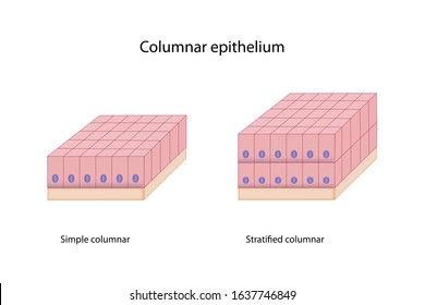
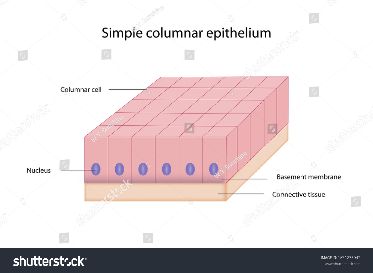
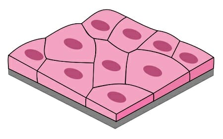
39 simple columnar epithelium diagram
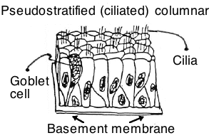
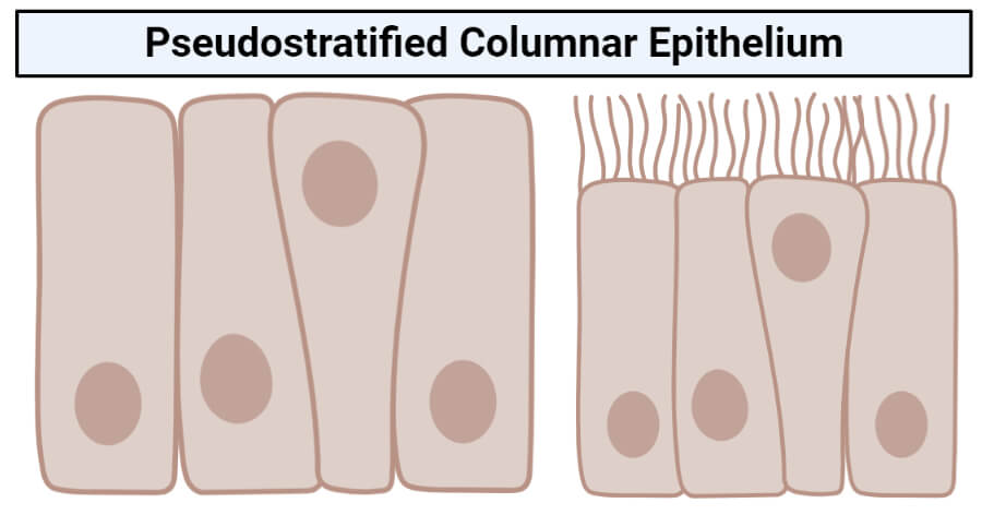
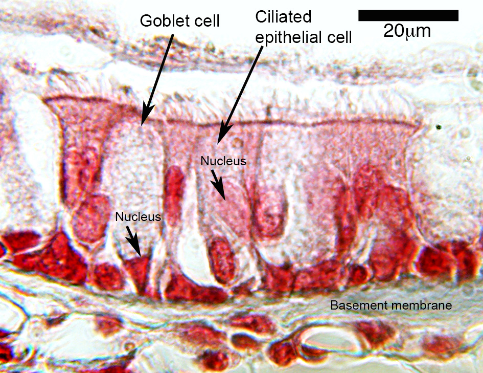
Simple columnar epithelium Illustrations Simple columnar epithelium Illustrations from Motifolio. Title: Simple columnar epithelium Keywords: Simple columnar epithelium illustration figure drawing diagram image This illustration is included in the following Illustration Toolkit Pseudostratified Columnar Epithelium under a Microscope ... The pseudostratified columnar epithelium comprises a single layer of cells but seems to be multilayered. It is because different cellular heights and nuclei are also placed at a different levels. I will show you the pseudostratified columnar epithelium under a light microscope with its identifying points and labeled diagram.
PDF Week 1: Covering and Lining Epithelia These labelled diagrams should closely follow the current Science courses in histology, anatomy and ... EPITHELIUM: simple columnar or pseudostratified columnar TISSUE / ORGAN: seminal vesicle (ox) 1 2 blood vessel basal cell basement membrane ciliated columnar cell lamina propria
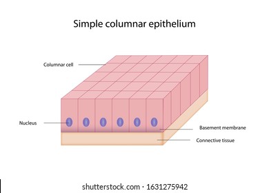
Simple columnar epithelium diagram
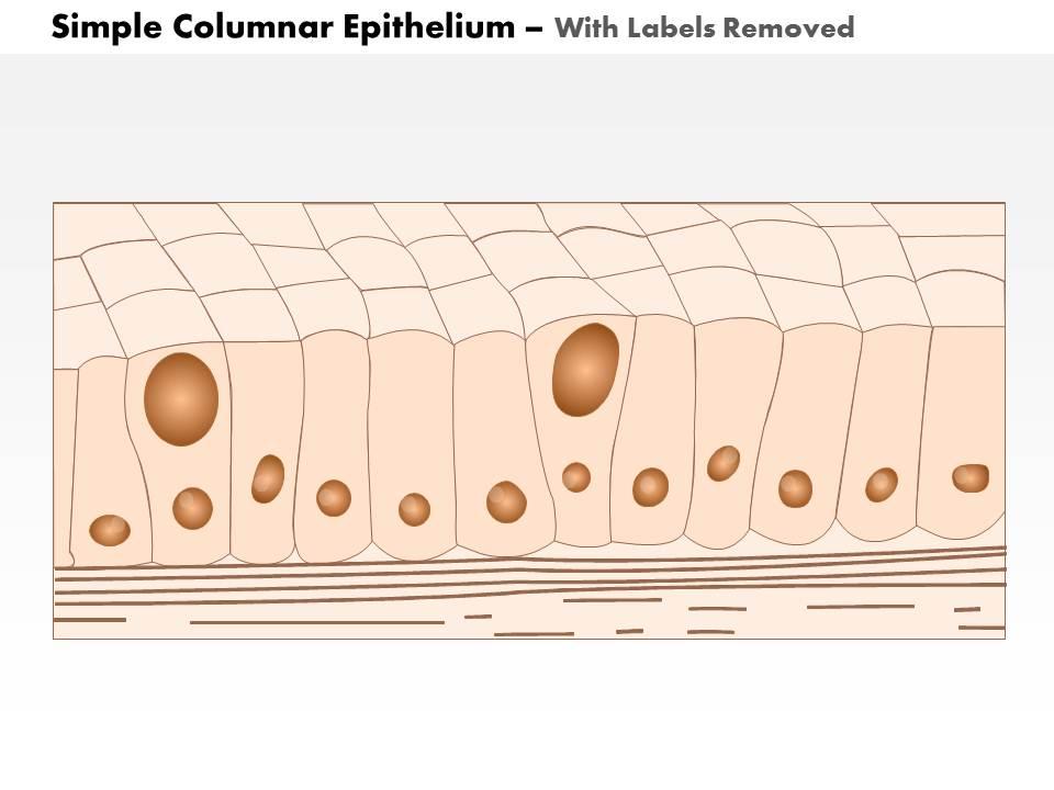
Simple columnar epithelium- structure, functions, examples Structure of the simple columnar epithelium The simple columnar epithelium is made up of a single layer of elongated cells which are always taller than they are wide and are directly attached to the basement membrane at the distal end. The nucleus is oval, large and is present at the base of the cell. Functions of Transitional Epithelium Tissue - Video ... 03.11.2021 · Transitional epithelium tissue is the somewhat self-explanatory name of a group of cells that can undergo a change in shape and composition. … Simple columnar epithelium - Austin Community College District Simple columnar epithelium (100X) Primate small intestine. At 100X you can begin to see the layer of simple columnar epithelium. the best place to look for it is on the surface of the villi. You can see what look like round white bubbles in the epithelium. These are goblet cells and they secrete mucus. The area inside the box is the portion of ...
Simple columnar epithelium diagram. 42 stratified cuboidal epithelium diagram - Modern Wiring ... Simple Columnar Epithelium Labeled Diagram Squamous. stratified squamous diagram photo of endothelial cells. Squamous means scale-like. simple squamous. Bodytomy provides a labeled diagram to help you understand the structure and Simple Columnar Epithelium: Labeled Diagram and Function. Epithelium is a tissue that lines the internal surface of ... Simple Cuboidal Epithelium Function & Location | What Is ... Simple Cuboidal Epithelium: Labeled Diagram Simple cuboidal epithelial cells are shaped like cubes, and the nucleus of each cell is large and located close to the center of the cell. Solved Examine the following: histologyguide.com 1. Simple ... Simple columnar epithelium MH119/120 (a) Draw a fully labelled captioned diagram at 40X showing the following: [7 +2] Columnar epithelial layer. Epithelial cells containing enterocytes and goblet cells, and Villi (finger-like projections) (4) (b) In what organ would you usually find this type of tissue? (1) This question hasn't been solved yet Ileum: Anatomy, histology, composition, functions - Kenhub The mucosa is lined by simple columnar epithelium (lamina epithelialis) comprising enterocytes and goblet cells. Underneath lies a connective tissue layer ( lamina propria) and a muscle layer ( lamina muscularis mucosae ). Compared to the rest of the small intestine the circular folds are rather flat and the villi relatively short.
Simple Columnar Epithelium: A Labeled Diagram and ... Let's understand the structure of this type of epithelium with the help of the labeled simple columnar epithelium diagram given below. The nucleus of each of the cells is usually placed quite close to the thin, sheet-like basement membrane. Most of the nuclei are placed at the same level. Epithelial Tissue | histology A. Simple columnar epithelium. Slide 29 (small intestine) View Virtual Slide Slide 176 40x (colon, H&E) View Virtual Slide Remember that epithelia line or cover surfaces. In slide 29 and slide 176, this type of epithelium lines the luminal (mucosal) surface of the small and large intestines, respectively. Refer to the diagram at the end of this chapter for the tissue orientation and consult ... Simple Columnar Epithelium Labeled Diagram Apr 20, 2019 · Simple Columnar Epithelium: A Labeled Diagram and Functions Epithelium is a tissue that lines the internal surface of the body, as well as the internal organs. Simple epithelium is one of the types of epithelium that is divided into simple columnar epithelium, simple squamous epithelium, and simple cuboidal epithelium. Simple Columnar Epithelium Diagram - Quizlet Start studying Simple Columnar Epithelium. Learn vocabulary, terms, and more with flashcards, games, and other study tools.
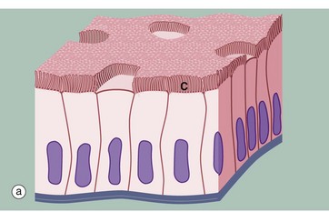
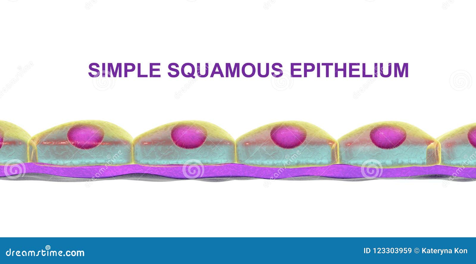
Simple Squamous Epithelium under a Microscope with a ... Simple squamous epithelium under a microscope consists of a single layer of thin, flat, and scale-like cells. These cells are joined together by an intercellular junction and rest on the basement membrane, whose thickness depends on the location. Here, I will show you what simple squamous looks like under a light microscope. Epithelial Tissue: Structure with Diagram, Function, Types ... Columnar- long or column-like cylindrical cells, which have nucleus present at the base On the basis of the number of layers present, epithelial tissue is divided into the simple epithelium and stratified or compound epithelium Simple Epithelium- it is composed of one layer of a cell and mostly has a secretory or an absorptive function Ciliated Epithelium - Concept, Structure, Function and ... A simple ciliated epithelium cell present in the pulmonary system is always interspersed with the goblet cells. It secretes mucus to form a mucosal layer apical to the epithelial layer. The rowing-like action of epithelial cilia always works in tandem with goblet cells to propel mucus that too away from the lungs. 4.2 Epithelial Tissue - Anatomy & Physiology Simple columnar epithelium forms a majority of the digestive tract and some parts of the female reproductive tract. Ciliated columnar epithelium is composed of simple columnar epithelial cells with cilia on their apical surfaces. These epithelial cells are found in the lining of the fallopian tubes where the assist in the passage of the egg ...
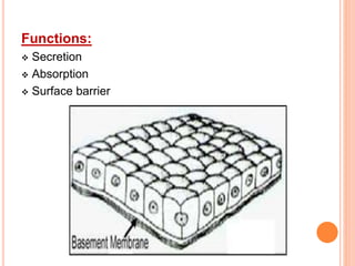
Simple Columnar Epithelium |Introduction ,Types, & Functions The Simple Columnar Epithelium is tissues composed of a single layer of long epithelial cells which are usually seen in an area where absorption and secretion are important facts. The cells of these epithelial are arranged in a neat row within nuclei at the same level, near to th basal end.. In a cross-section of organs, these cells look like thin columns, which differentiate them from ...
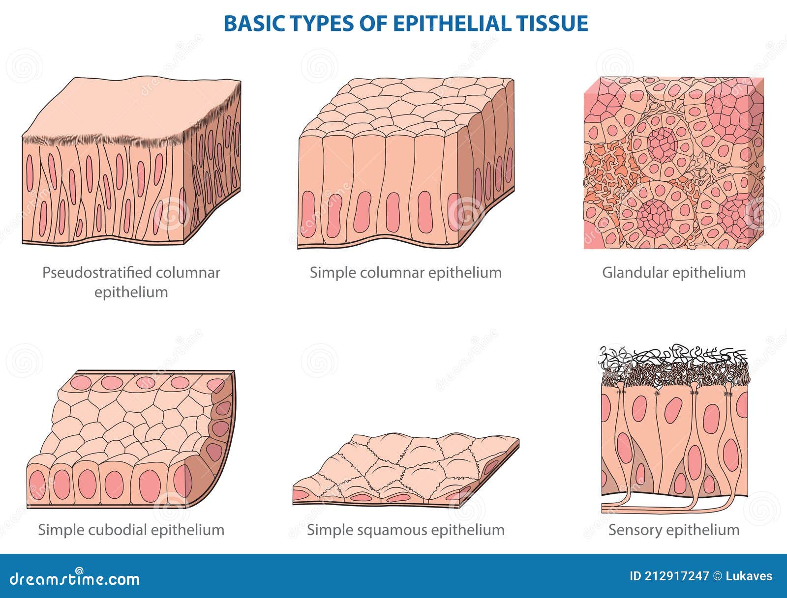
Epithelium - Wikipedia Epithelium / ˌ ɛ p ɪ ˈ θ iː l i ə m / is one of the four basic types of animal tissue, along with connective tissue, muscle tissue and nervous tissue.It is a thin, continuous, protective layer of compactly packed cells with little intercellular matrix.Epithelial tissues line the outer surfaces of organs and blood vessels throughout the body, as well as the inner surfaces of cavities in ...
Simple Columnar Epithelium - Definition & Function ... Simple Columnar Epithelium Definition Simple columnar epithelia are tissues made of a single layer of long epithelial cells that are often seen in regions where absorption and secretion are important features. The cells of this epithelium are arranged in a neat row with the nuclei at the same level, near the basal end.
Chapter 1, Page 10 - HistologyOLM - SteveGallik.org Diagram of a simple secretory columnar epithelium in section. A simple secretory columnar epithelium is found at two major sites in the animal body: 1) lining the stomach, and 2) lining the cervical canal. Our microscopic study of simple secretory columnar epithelium will consist of a study of the following specimen: Study #1.
Stomach histology: Mucosa, glands and layers | Kenhub 28.02.2022 · The surface mucous cells, also known as foveolar epithelium, are the simple columnar epithelium lining the lumen of the stomach. They secrete alkaline, highly viscous mucus, which closely adheres to the cellular surface. The mucus protects the stomach lining by minimising the abrasion from food particles and forming a physical barrier from the hydrochloric …
Solved Short answer questions 20. In the following ... In the following schematic diagram of a simple columnar epithelium lining the digestive tract, indicate which position (A to E) along the apical -basal axis best corresponds to each of the features below. (5 pts) Gut lumen Connective tissue (E) Basal lamina (A) Cell apical surface (B) Adherens junctions (D) Hemidesmosomes (C) Gap junctions 21.
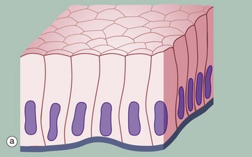
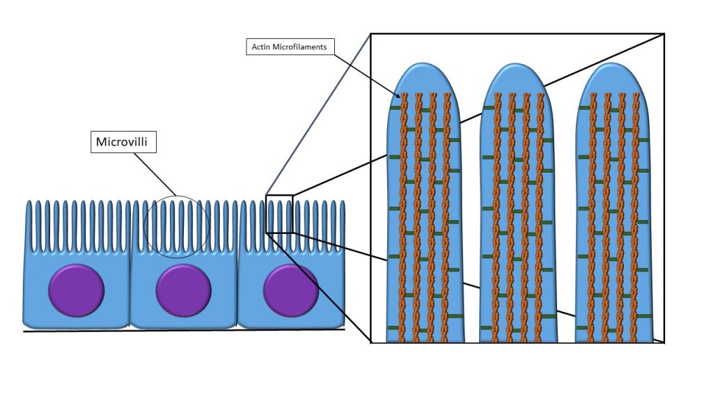
Simple epithelium: Location, function, structure | Kenhub Synonyms: Simple columnar epithelium (with microvillous border) In this type of epithelium, the height of cells exceeds the width of the cell and seem closely packed narrow columns. The apical surfaces of these cells face the lumen and the opposite surface faces the basement membrane. The ovoid nuclei are usually placed towards basal surface.
Simple Columnar Epithelium Diagram | Quizlet Start studying Simple Columnar Epithelium. Learn vocabulary, terms, and more with flashcards, games, and other study tools.
Simple Squamous Epithelium: Location and Diagram - Video ... The simple squamous epithelium location specifically exists in the lining of the blood vessels like the arteries, veins, and capillaries. It is also found lining the alveoli or air sacs within the...
Simple Columnar Epithelium Labeled Diagram Simple Columnar Epithelium Labeled Diagram Ciliated columnar epithelium is composed of simple columnar epithelial cells with cilia on their apical This illustration shows a diagram of a goblet cell. These labelled diagrams should closely follow the current Science (simple squamous epithelium). ORIGIN: columnar epithelium with goblet cells. TISSUE .
Simple columnar epithelium - Austin Community College District Simple columnar epithelium (100X) Primate small intestine. At 100X you can begin to see the layer of simple columnar epithelium. the best place to look for it is on the surface of the villi. You can see what look like round white bubbles in the epithelium. These are goblet cells and they secrete mucus. The area inside the box is the portion of ...
Functions of Transitional Epithelium Tissue - Video ... 03.11.2021 · Transitional epithelium tissue is the somewhat self-explanatory name of a group of cells that can undergo a change in shape and composition. …
Simple columnar epithelium- structure, functions, examples Structure of the simple columnar epithelium The simple columnar epithelium is made up of a single layer of elongated cells which are always taller than they are wide and are directly attached to the basement membrane at the distal end. The nucleus is oval, large and is present at the base of the cell.








:background_color(FFFFFF):format(jpeg)/images/article/en/simple-epithelium/VKbSAUnFnI6qqyq2cS0ag_qDDI8y5Bsv32llNotfSCA_Simple_columnar_epithelium_with_striated_border01.png)





0 Response to "39 simple columnar epithelium diagram"
Post a Comment