41 diagram of a prokaryotic cell
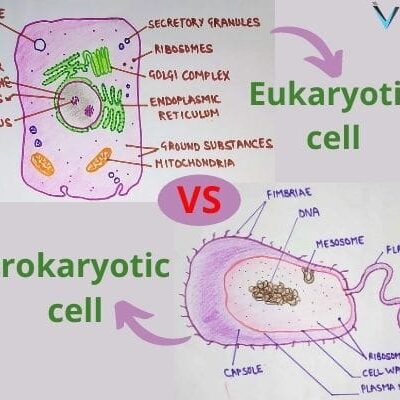
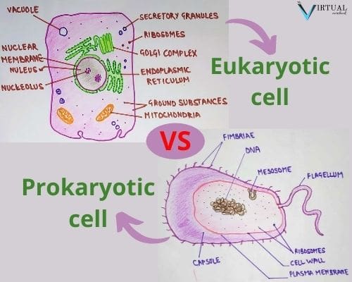
Prokaryotes and Eukaryotes Venn Diagram Prokaryotes Both Prokaryotes Eukaryotes and Eukaryotes *No Nucleus *Cells have a nucleus *Small and simple *Cells have organelles *No organelles *Can be unicellular or *Are very abundant *Have ribosomes multicellular *All are unicellular *Have DNA *Have a cytoskeleton ... Prokaryotic Cell Diagram. to help you remember prokaryotes parts and pieces. - fimbriae: allow bacteria to adhere to target host cells, and play a major role in bacterial virulence. - conjugation pili: the tubes used to transfer plasmids from donor to recipient bacteria. This article has.
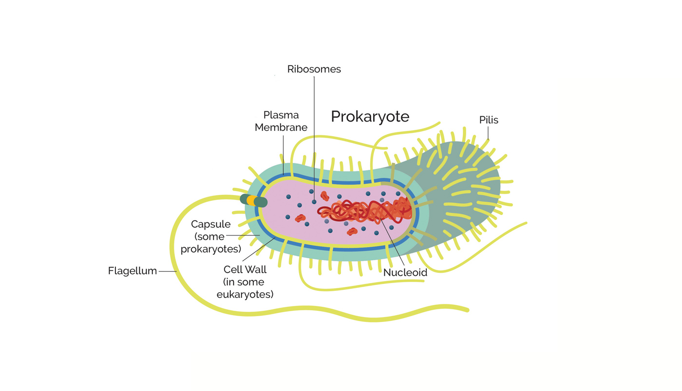
You are expected to be able to annotate a diagram you draw of a prokaryotic cell with the following functions:Plasma membrane - controls the ...

Diagram of a prokaryotic cell
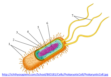
A rigid structure that surrounds the cell membrane and provides support to the cell plasma membrane A selectively-permeable phospholipid bilayer forming the boundary of the cells Flagellum A long, hairlike structure that grows out of a cell and enables the cell to move. hook ... basal body anchors the cilium or flagellum peptidoglycan 3. Two major categories of cells Prokaryotic cell: simple internal structure cell flagella Eukaryotic cell: complex internal structure Prokaryotic cells (bacteria, archaea) Eukaryotic (plants, animals, fungi, protists) When you need to see a cellular tower location map to find your nearest cell tower, there are a few options, as shown by Wilson Amplifiers. You can use a website or smartphone app to find the nearest tower for cellular service, or you can c...
Diagram of a prokaryotic cell. Prokaryotic cell structure diagram, vector illustration cross section labeled scheme. Microbiology science educational information. Micro organism research and bacteria study. Cell elements example. Formats EPS 3000 × 2500 pixels • 10 × 8.3 in • DPI 300 • JPG Contributor V VectorMine Similar images See all Assets from the same collection See all Prokaryotic Cells Eukaryotic Cells . Title: Prokaryotic and Eukaryotic Venn Diagram Worksheet Author: Brandon Linn Created Date: 9/19/2018 6:59:56 PM ... The Cell Wall of Prokaryotes. The cytoplasm of prokaryotic cells has a high concentration of dissolved solutes. Therefore, the osmotic pressure within the cell is relatively high. The cell wall is a protective layer that surrounds some cells and gives them shape and rigidity. Cell wall and glycocalyx • Not all cells have cell wall • Simpler cell wall construction than in prokaryotes •Cellulose - Most algae, plants, some fungi (chitin) • Polysaccharides glucan and mannan -yeast • Pellicle (not cell wall, atypical covering) -protozoans • Glycocalyx - Sugar coating - Increases cell strength ...
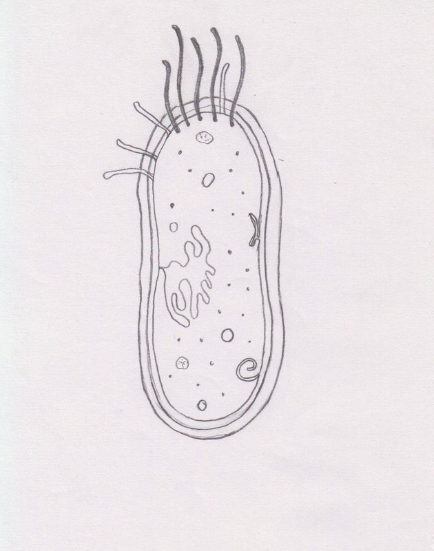
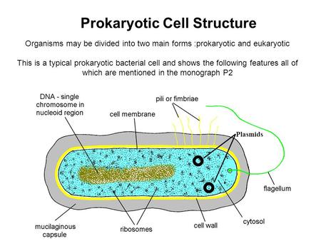
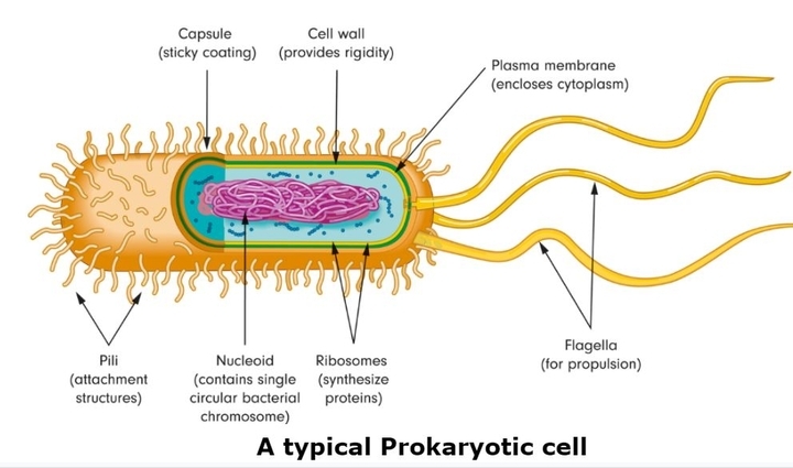
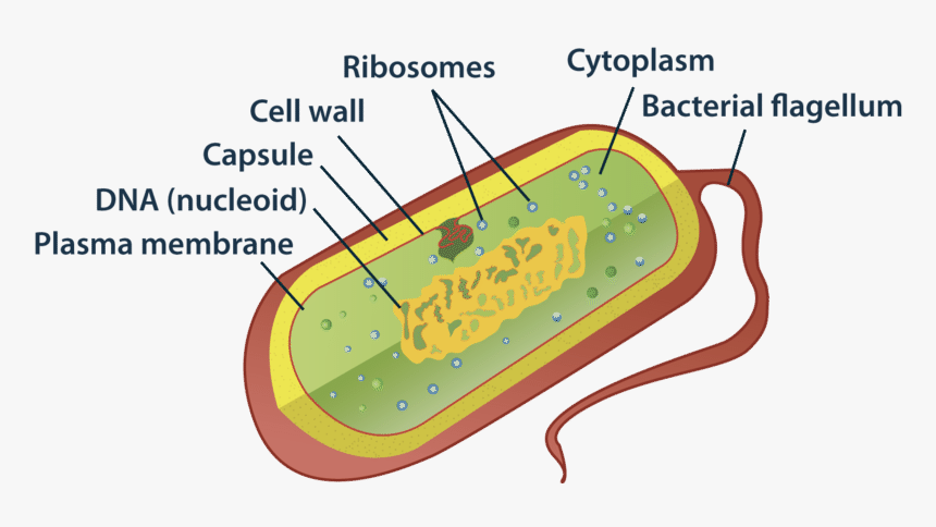
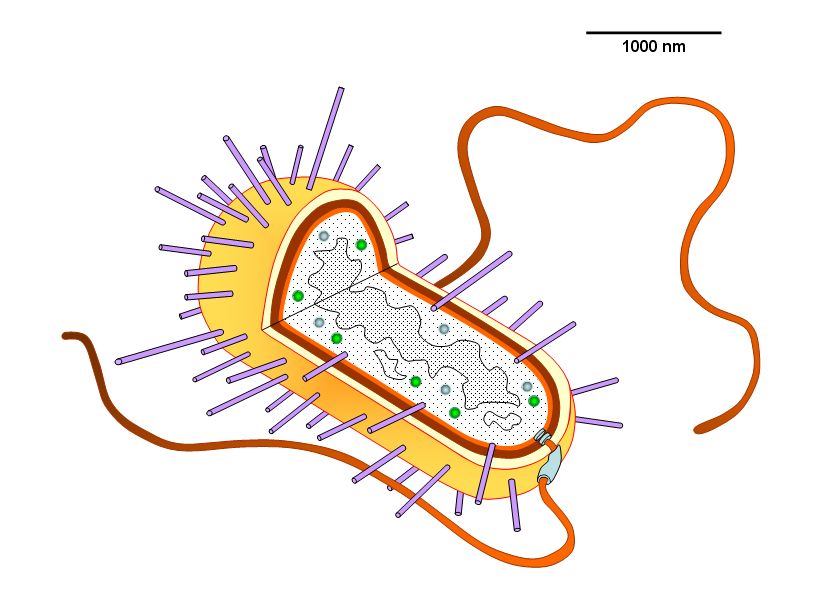
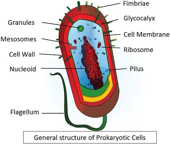
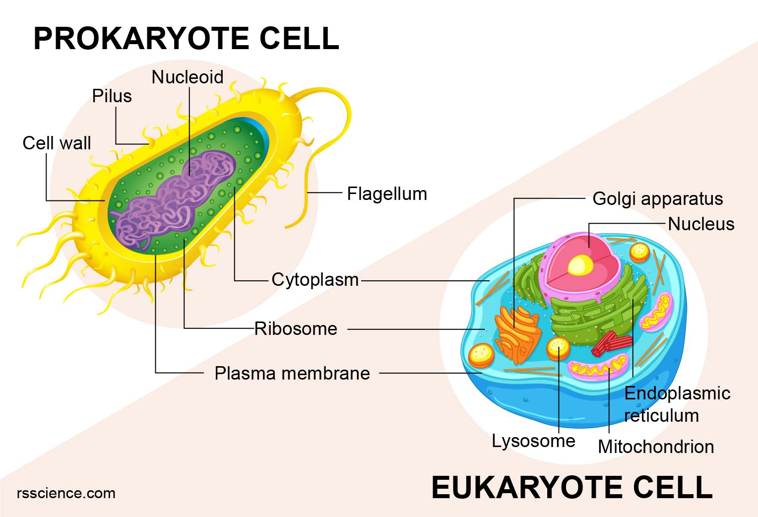
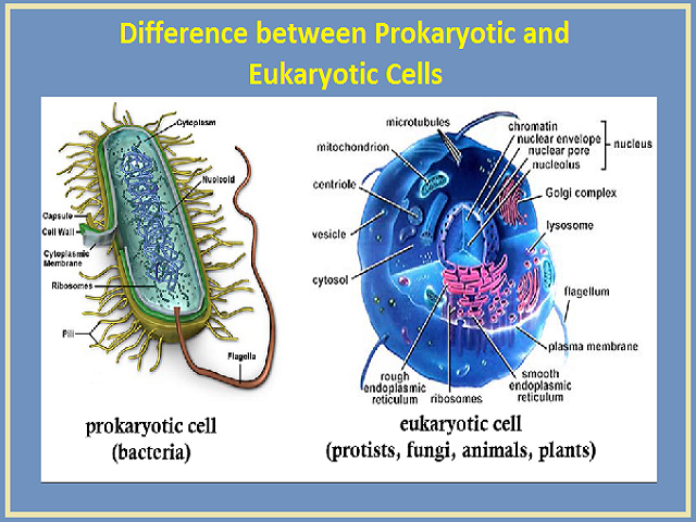
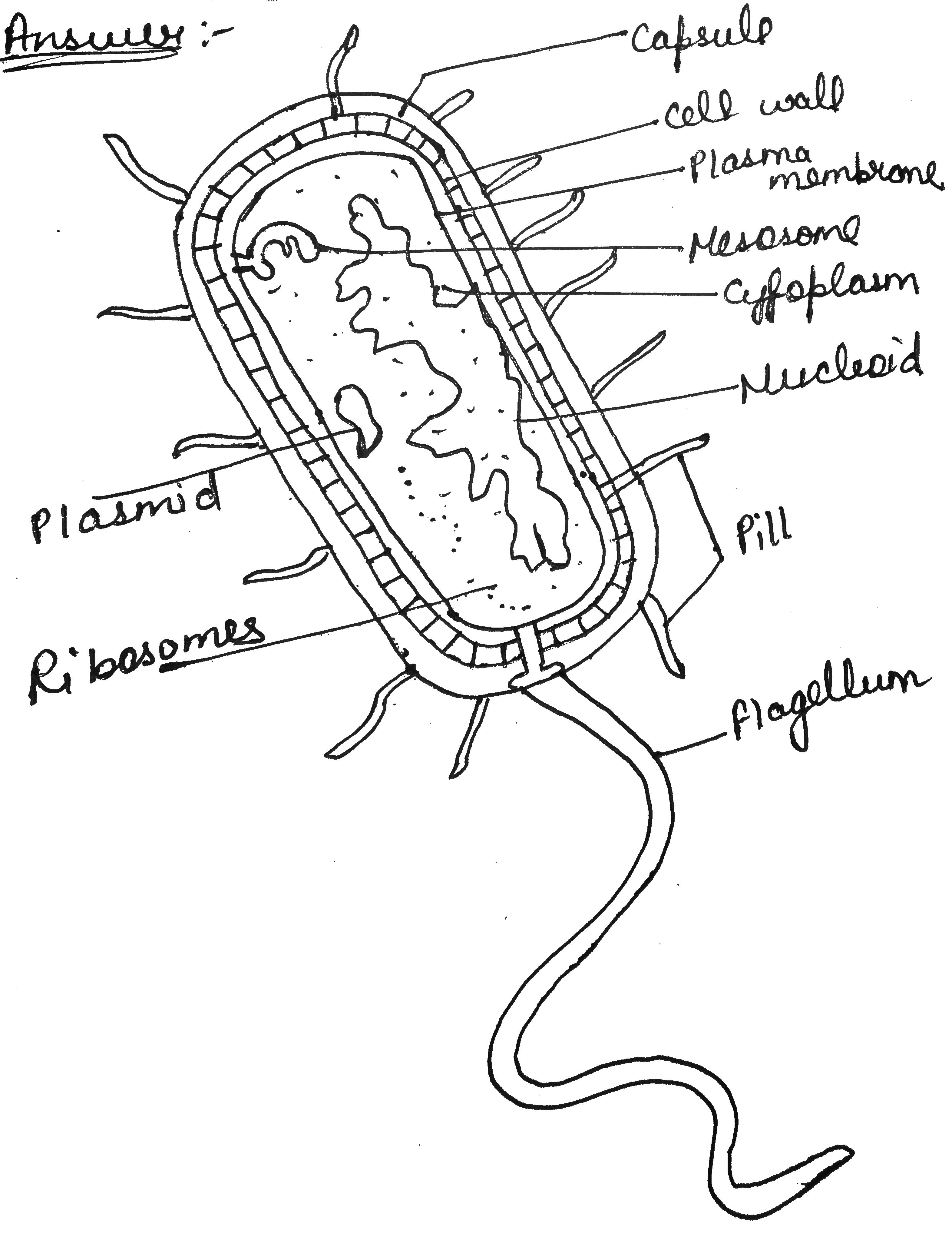
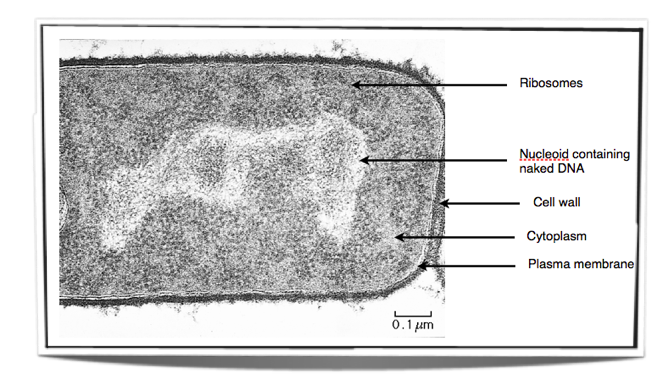
Prokaryotic Cell Definition: A prokaryotic mobile is one that lacks a true nucleus and membrane-sure organelles. micro organism and Archaea organisms are based at the prokaryotic cell, while all different styles of lifestyles are eukaryotic. however, organisms with prokaryotic cells are considerable and account for a huge portion of the Earth’s biomass. Features of Prokaryotic Cells The genetic material is localized in a region known as nucleoid and it has no surrounding membrane. These cells contain large numbers of the ribosome for protein synthesis In some prokaryotes, the cell membrane folds to form structures known as mesosomes which assist in cell respiration. Diagram of a Prokaryotic Cell Prokaryotic Cell Components The four main components of prokaryotic cells are: Plasma Membrane- The plasma membrane is a protective layer of phospholipid molecules that isolates the cell from its surroundings. Cytoplasm- Inside the cell, there is a jelly-like fluid called cytoplasm. Dec 18, 2016 · Prokaryotic Cell Diagram. The following image is a diagram of a prokaryotic cell; in this case, a bacterium. The Anatomy of a Bacterial Cell Prokaryotic Cell Structure. Prokaryotic cells do not have a true nucleus that contains their genetic material as eukaryotic cells do.
Cell size. Typical prokaryotic cells range from 0.1 to 5.0 micrometers (μm) in diameter and are significantly smaller than eukaryotic cells, which usually have diameters ranging from 10 to 100 μm. The figure below shows the sizes of prokaryotic, bacterial, and eukaryotic, plant and animal, cells as well as other molecules and organisms on a ... 05.06.2021 ... Unicellular organisms of the domains Archaea and Bacteria are classified as prokaryotes. Prokaryotic cells lack membrane-bound cellular ... Whereas eukaryotic cells have many different functional compartments, divided by membranes, prokaryotes only have one membrane (the plasma membrane) ... Prokaryotes-CellShapes Most bacteria are classifies according to shape: 1. bacillus (pl. bacilli)= rod-shaped 2. coccus(pl. cocci… sounds like cox-eye)= spherical 3. spiral shaped a. spirillum(pl. spirilla) = spiral with rigid cell wall, flagella b. spirochete(pl. spirochetes)= spiral with flexible cell wall, axial filament
Introduction to Prokaryotes, Eukaryotes
Skin cancer is the most common type of cancer in the United States, with Basal and Squamous Cell skin cancer being the most common carcinoma types. There are roughly 5.4 million diagnoses of these two types every year. Basal Cell Carcinoma ...

Eukaryotic and Prokaryotic Cells - The Science and Maths Zone
Basic Cell Types: Prokaryotic are cells that lack a nucleus (nuclear membrane). Prokarotic cells are single cells but are subdivided into Bacteria and Arachaea as mention in the previous slide. Eukaryotic cells contain a nucleus (nuclear membrane). Eukaryotic cells include: plants, animals, fungi and protists ( a

Prokaryotes are the organisms which have primitive nucleus ...
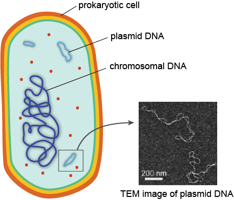
Prokaryotic cells are not as complex as eukaryotic cells. They have no true nucleus as the DNA is not contained within a membrane or separated from the rest of the cell, but is coiled up in a region of the cytoplasm called the nucleoid. Prokaryotic organisms have varying cell shapes.
2.2 Prokaryotic cell - BIOLOGY4IBDP
File:Prokaryote cell diagram.svg. Size of this PNG preview of this SVG file: 800 × 441 pixels. Other resolutions: 320 × 177 pixels | 640 × 353 pixels | 1,024 × 565 pixels | 1,280 × 706 pixels | 2,560 × 1,412 pixels | 870 × 480 pixels. This is a file from the Wikimedia Commons. Information from its description page there is shown below.

Lecture 4 Prokaryote Cell Structure & Function Cell Diagram ...
Click here to get an answer to your question ✍️ Draw a diagram of a prokaryotic cell and label at least four parts in it.
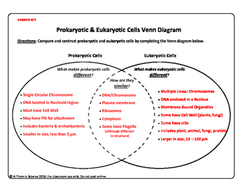
Prokaryotic Cell versus Eukaryotic Cell Venn Diagram
Animal and plant cells are types of eukaryotic cells, whereas bacteria are a type of prokaryote; Prokaryotic cells are much smaller than eukaryotic cells (between 100 - 1000 times smaller); They also differ from eukaryotic cells in having: A cytoplasm that lacks membrane-bound organelles; Their ribosomes are structurally smaller (70 S) in comparison to those found in eukaryotic cells (80 S)
2.2 Prokaryotic Cells. - ppt download
Prokaryotic Cell Diagram Prokaryotic Cell Structure All prokaryotic cell shares four common components:- 1. Plasma membrane - It refers to the outer membrane which separates the inner environment from the external environment. It is a thin lipid bilayer. It is selectively permeable. 2.

ultrastructure
Prokaryotic cells have various shapes; the four basic shapes of bacteria are: Cocci - A bacterium that is spherical or ovoid is called a coccus (Plural, cocci). e.g. Streptococcus, Staphylococcus. Bacilli - A bacterium with cylindrical shape called rod or a bacillus (Plural, bacilli).

Cell Structure (Eukaryotic vs Prokaryotic) Diagram | Quizlet
Many prokaryotic cells have sphere, rod, or spiral shapes (as shown below). In the following sections, we'll walk through the structure of a prokaryotic cell, starting on the outside and moving towards the inside of the cell. _Image modified from " Bacterial morphology diagram ," by Mariana Ruiz Villareal (public domain)._ The capsule
Prokaryotic Cells - Definition, Structure, Characteristics ...
Prokaryotic Cell Diagram to help you remember prokaryotes parts and pieces. Cytoskeleton: It's a relatively recent scientific discovery that rod-shaped bacteria and Archaea possess cytoskeletal proteins that function similarly to the cytoskeleton of eukaryotic cells.
Draw A Neat Labelled Diagram Of Prokaryotic Cell, HD Png ...
The prokaryotic cell diagram given below represents a bacterial cell. It depicts the absence of a true nucleus and the presence of a flagellum that differentiates it from a eukaryotic cell. Prokaryotic Cell Diagram illustrates the absence of a true nucleus Components of Prokaryotic Cells The prokaryotic cells have four main components:

Prokaryotic Cell and Eukaryotic Cell
16.11.2021 ... Prokaryotic cells (Prokaryotes) are cells without a true nucleus and lack membrane-bound organelles. The organisms are known as Prokaryotes.

Structure of a bacterial cell. Anatomy of the prokaryote ...
A non-membrane-bounded region in a prokaryotic cell where the DNA is concentrated. cytoplasm A jellylike fluid inside the cell in which the organelles are suspended capsule Covers the cell wall in prokaryotes. cytoplasmic membrane a semipermeable barrier that separates the cell interior (cytoplasm) from the environment cell wall
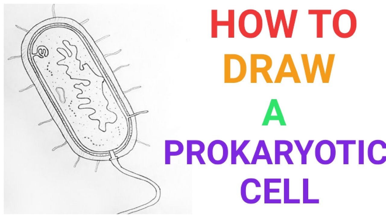
How to draw a prokaryotic cell | prokaryotic organism | Bacterial cell | Easily | Well lebelled diag
Feb 26, 2014 - The typical textbook diagram of a prokaryotic cell. The focus here is on structures rather than functions and the interior of this cell ...

BIO 149 Prokaryotic Cell Diagram Diagram | Quizlet
Prokaryotes. Bacteria are amongst the simplest of organisms - they are made of single cells. Their cell structure is simpler than the cells of eukaryotes and cells are smaller, most are 0.2 μm ...

Diagram of Prokaryotic Cell
Nov 19, 2021 · The prokaryotic cell diagram given below represents a bacterial cell. At the end of class students will be given a blank Venn diagram and asked to fill it in with the similarities and differences of prokaryotic and eukaryotic cells. Unlabeled animal cell diagram. Quiz yourself by filling in the blanks.
320 × 192 Pixels - Prokaryotic Cell Diagram Transparent PNG ...
The internet is your best friend when looking up cell phone numbers. There are a variety of sites and tools that make it simple to perform a cell phone number search. Some tools are free to use while others may charge a nominal fee. Simply ...
Unlabeled prokaryotic cell diagram free image download
Eukaryotic Cell Vs Prokaryotic Cell Difference And Comparison Diffen Prokaryotic Cell Eukaryotic Cell Prokaryotes. Prokaryotic And Eukaryotic Cells Venn Diagram Activity In 2021 Eukaryotic Cell Venn Diagram Venn Diagram Activities. Prokaryotic Cell Versus Eukaryotic Cell Venn Diagram Eukaryotic Cell Prokaryotic Cell Cells Worksheet.
What's the difference between prokaryotic and eukaryotic ...
Structure of a prokaryotic cell. The prokaryotic cells is not so complex as cells of eukaryotic origin because they are cellular organelles. Most prokaryotic cells contain the following componentsor parts: Capsule. This is an extra external covering found in certain prokaryotic cells, which serves to guard the cells against foreign invaders.
Difference Between Prokaryotic Cells and Eukaryotic Cells ...
... a basic diagram on the elements conforming a prokaryote cell. in this case a bacteria. Date, 26 April 2006. Source, i did the diagram myself using adobe ...
PROKARYOTES AND EUKARYOTES - The Virtual Notebook
Prokaryotic Cell Diagram The prokaryotic cell diagram given below represents a bacterial cell. Made of single cell. It depicts the absence of a true nucleus and the presence of a flagellum that differentiates it from a eukaryotic cell. The Anatomy of a Bacterial Cell.

A schematic diagram of a prokaryotic cell. Source: [1 ...
When you need to see a cellular tower location map to find your nearest cell tower, there are a few options, as shown by Wilson Amplifiers. You can use a website or smartphone app to find the nearest tower for cellular service, or you can c...

Biology 101: Cells - Owlcation
3. Two major categories of cells Prokaryotic cell: simple internal structure cell flagella Eukaryotic cell: complex internal structure Prokaryotic cells (bacteria, archaea) Eukaryotic (plants, animals, fungi, protists)

Prokaryotic Cell Diagram | Quizlet
A rigid structure that surrounds the cell membrane and provides support to the cell plasma membrane A selectively-permeable phospholipid bilayer forming the boundary of the cells Flagellum A long, hairlike structure that grows out of a cell and enables the cell to move. hook ... basal body anchors the cilium or flagellum peptidoglycan
Eukaryotes and Prokaryotes - What are the Similarities ...
Prokaryotes vs Eukaryotes: What Are the Key Differences ...
What is the difference between Prokaryotic and Eukaryotic Cells?
Draw a well-labelled diagram of a prokaryoticcell. | Snapsolve
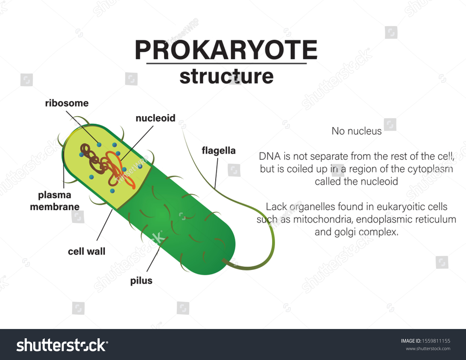
Diagram Structure Prokaryotic Cell Stock Vector (Royalty Free ...

Prokaryotes vs Eukaryotes- Definition, 47 Differences, Examples
IB Biology Notes - 2.2 Prokaryotic cells
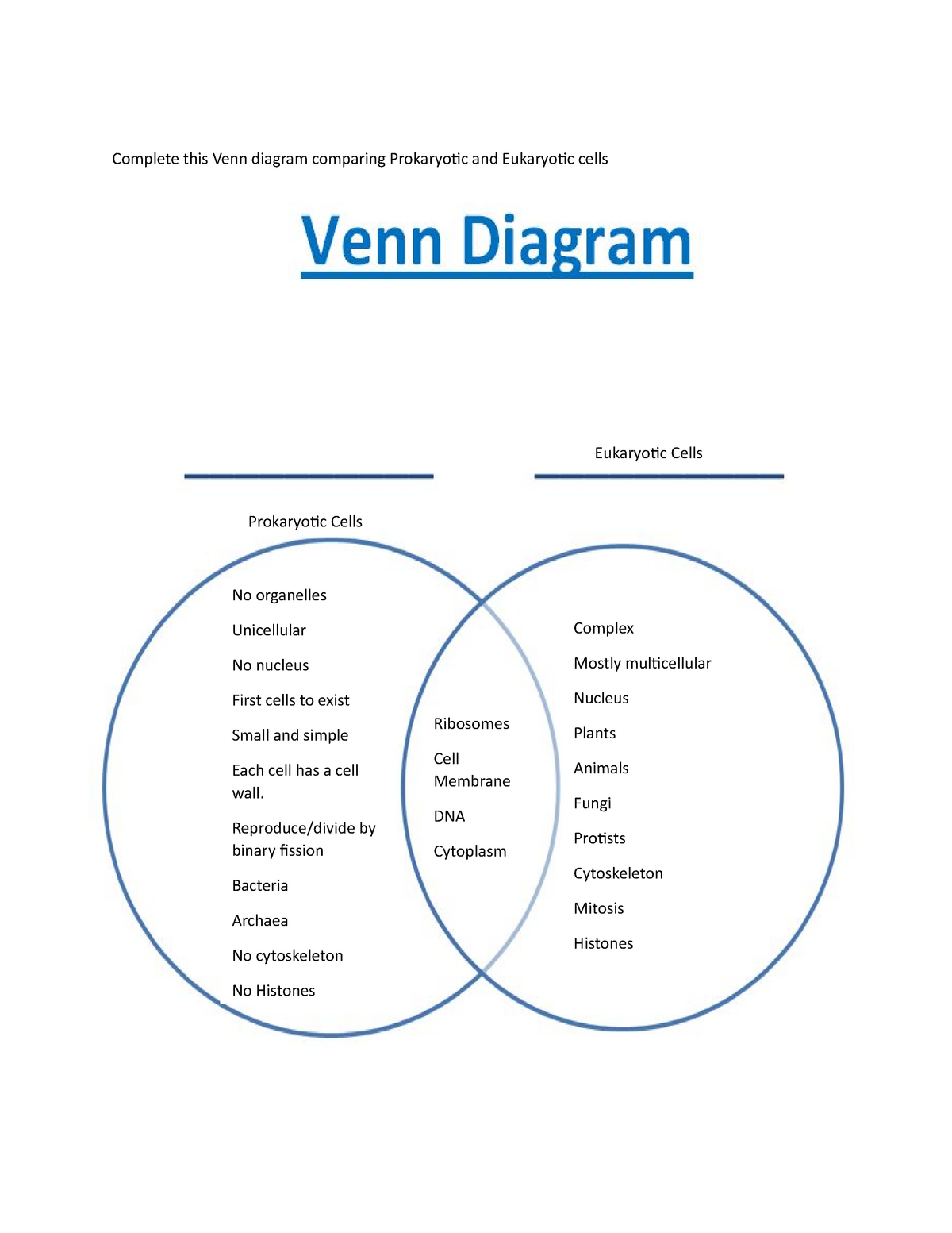
Venn diagram student handout - Complete this Venn diagram ...

Structure of Prokaryotic Cell | Notes, Videos, QA and Tests ...
Cells - Prokaryotic Cell Structure and Function | Shmoop

72,560 Prokaryotic Cell Photos and Premium High Res Pictures ...
Difference between prokaryotic and eukaryotic cell - The ...
2.2 Prokaryotic cell - BIOLOGY4IBDP

Prokaryotic Cells | BioNinja

Review for Cell Test
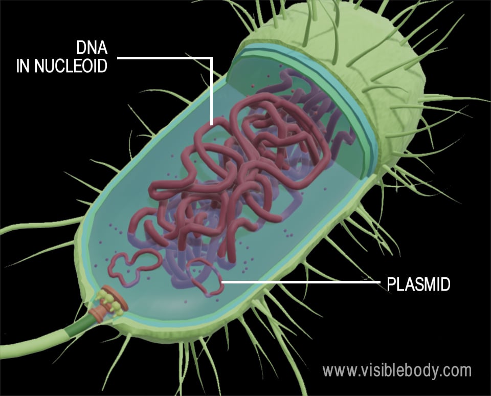
Prokaryotic Chromosomes
Draw a neat labelled diagram of a prokaryotic cell ...
0 Response to "41 diagram of a prokaryotic cell"
Post a Comment