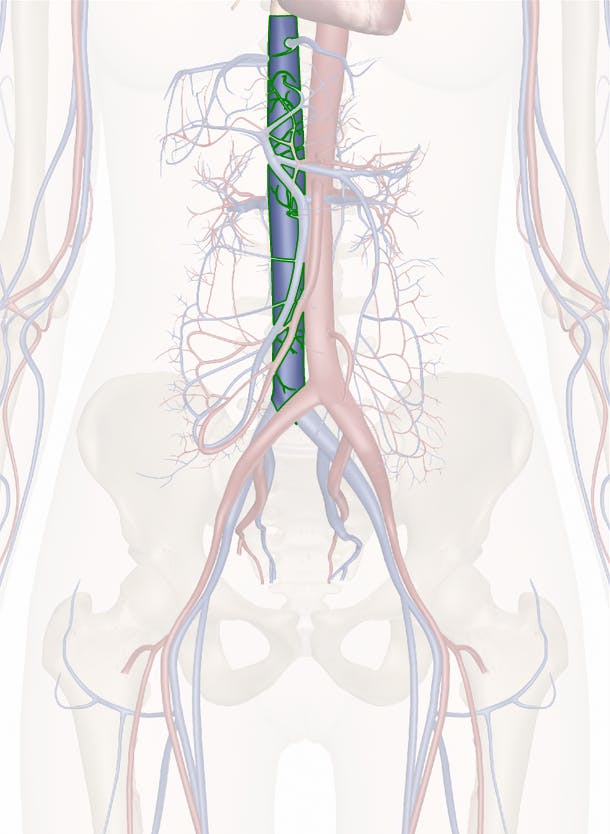
41 inferior vena cava diagram
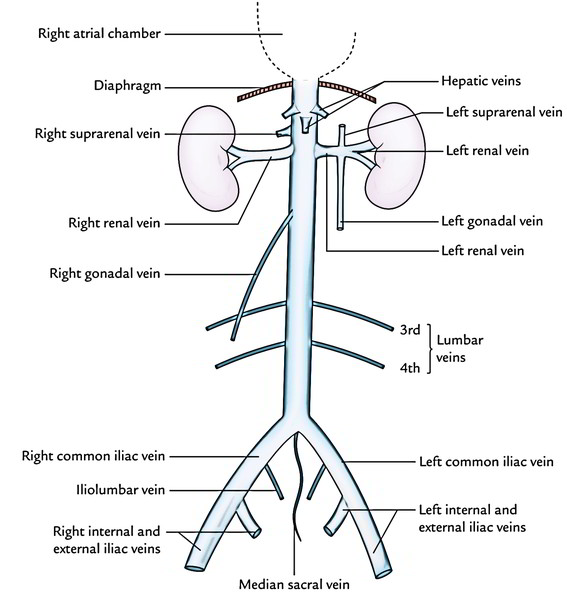
Inferior Vena Cava Diagram - Quizlet Start studying Inferior Vena Cava. Learn vocabulary, terms, and more with flashcards, games, and other study tools. Surgical anatomy of the hepatic veins and the inferior ... Accurate knowledge of the surgical anatomy of the hepatic veins and inferior vena cava is indispensable for surgeons, since catastrophes usually occur in this area. Material from 83 autopsies was examined concerning the patterns of ramifications of the hepatic veins along with right suprarenal and i …
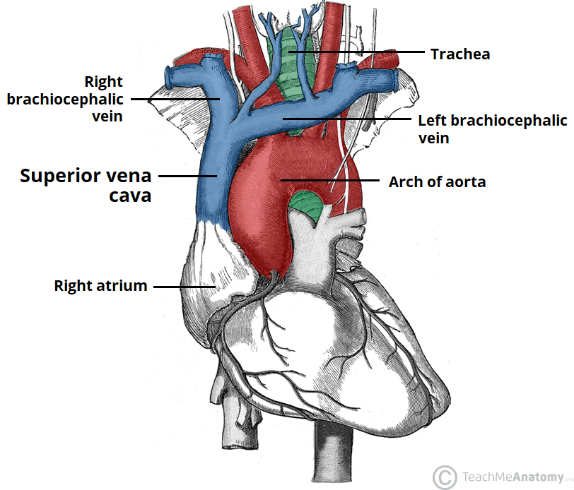
Dissection of the Sheep Heart - Houston Community College Locate the inferior vena cava and superior vena cava. 7. Use the scalpel to open the heart chambers: make an incision along the coronal plane, from the superior portion of the left ventricle to the superior portion of the right ventricle. Inferior Vena Cava Superior Vena Cava Posterior View Vena Cava = Singular Vena Cavae = Plural NOTICE the ...

Inferior vena cava diagram
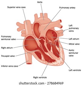
Inferior Vena Cava: Anatomy, Function, and Significance Apr 01, 2022 · The inferior vena cava (also known as IVC or the posterior vena cava) is a large vein that carries blood from the torso and lower body to the right side of the heart. From there the blood is pumped to the lungs to get oxygen before going to the left side of the heart to be pumped back out to the body. Schematic of inferior vena cava measurements during ... Schematic of inferior vena cava measurements during expiration (left) and inspiration (right). Short-axis measurements obtained after a 90 rotation of the transducer approximate the level of... Label the Heart - Labelled diagram Label the Heart - Labelled diagram. Superior Vena Cava, Aorta, Inferior Vena Cava, Pulmonary Vein, Pulmonary Artery.
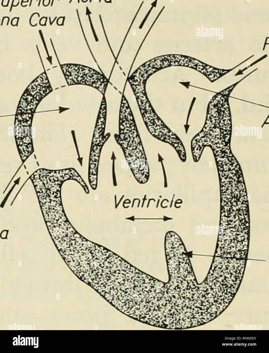
Inferior vena cava diagram. Blood Flow: Diagram - THE SYSTEMIC CIRCULATION Right ... Inferior Vena Cava drains venous blood from the lower regions of. Superior Vena Cava drains venous blood from the upper regions of. Aorta Ascending aorta delivers blood to heart neck, head, and arms; Descending aorta delivers blood to visceral and lower body tissues. THE CORONARY CIRCULATION. Ascending Aorta deliv ers blood to heart neck, /10 Heart Labels Part A: Label the heart diagram | Chegg.com Transcribed image text: /10 Heart Labels Part A: Label the heart diagram below using the following structure names: left/right atrium, left/right ventricle, interventricular septum, myocardium, aorta, inferior vena cava, superior vena cava, pulmonary vein, pulmonary arteries, aortic valve, mitral (bicuspid) valve, pulmonary valve, tricuspid valve, Av bundle, AV node, bundle branches, Purkinje ... Duplication of inferior vena cava | Radiology Reference ... Pathology The inferior vena cava has a convoluted development during the 7-10 th weeks of gestation 4. posterior cardinal vein appears first but forms only the distal IVC i.e. iliac bifurcation. subcardinal veins (2) appear next, left subcardinal vein regresses, and right subcardinal vein forms the suprarenal IVC. Inferior Vena Cava - Anatomy Pictures and Information Dec 15, 2016 · The inferior vena cava is the largest vein in the human body. It collects blood from veins serving the tissues inferior to the heart and returns this blood to the right atrium of the heart. Although the vena cava is very large in diameter, its walls are incredibly thin due to the low pressure exerted by venous blood.
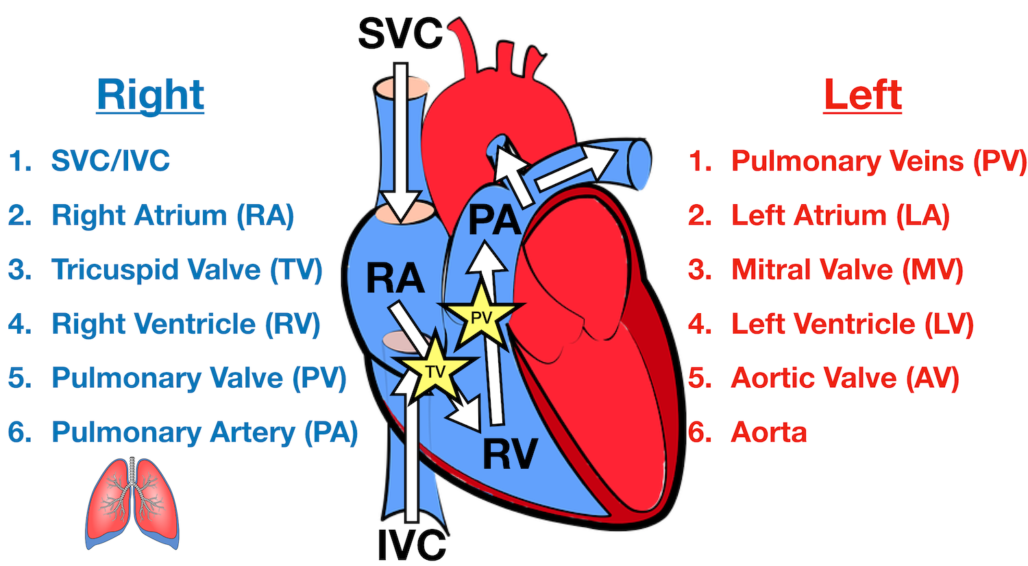
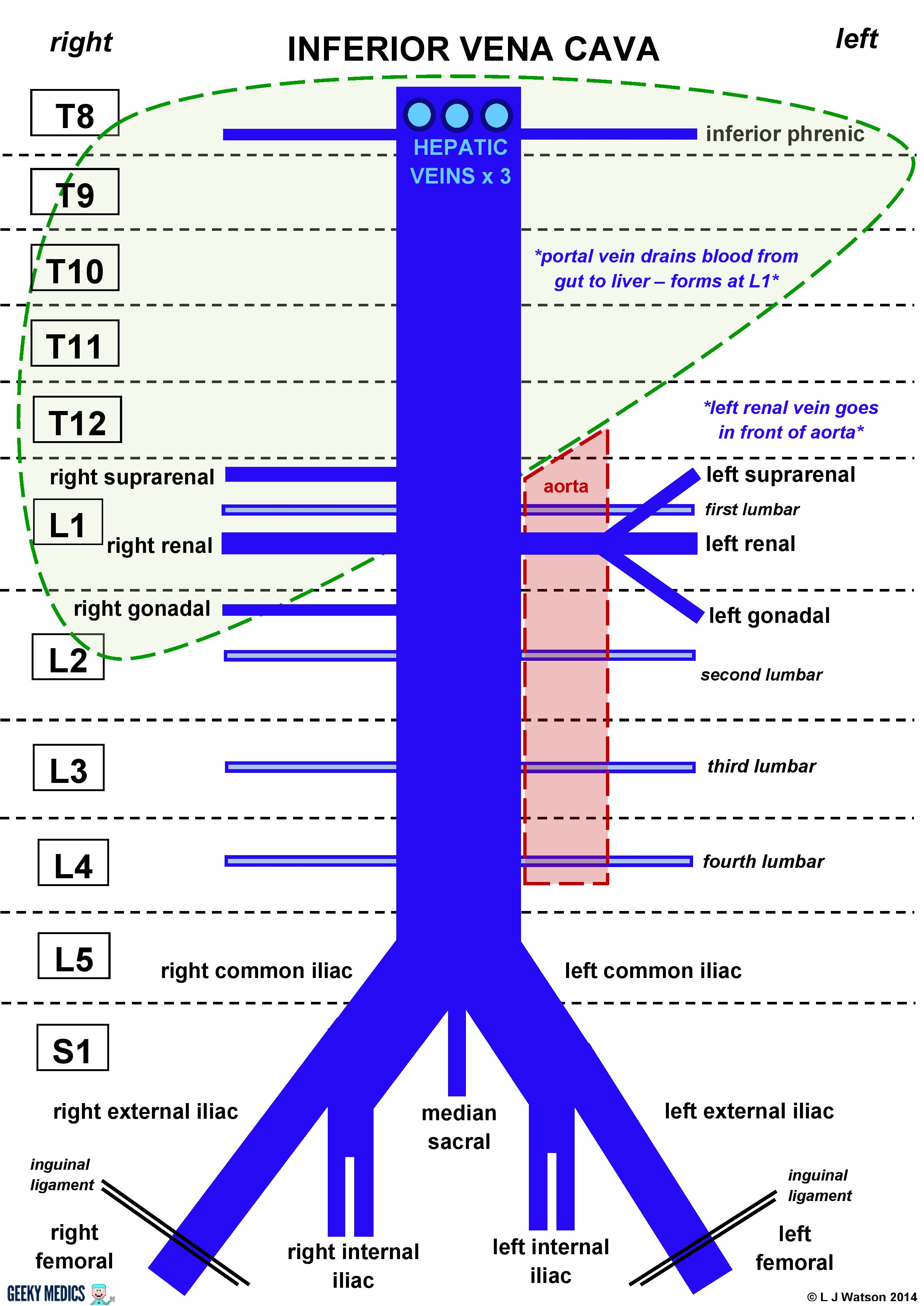
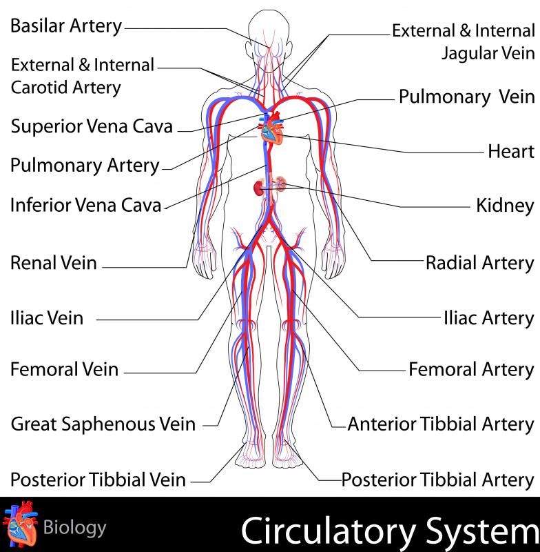
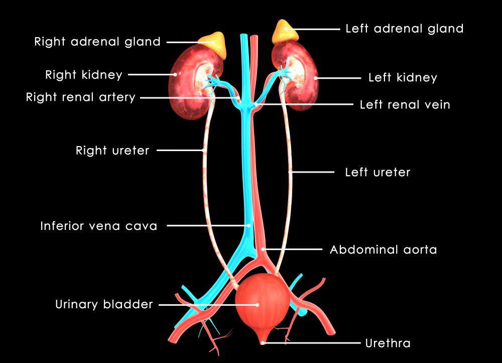
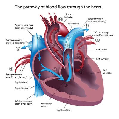
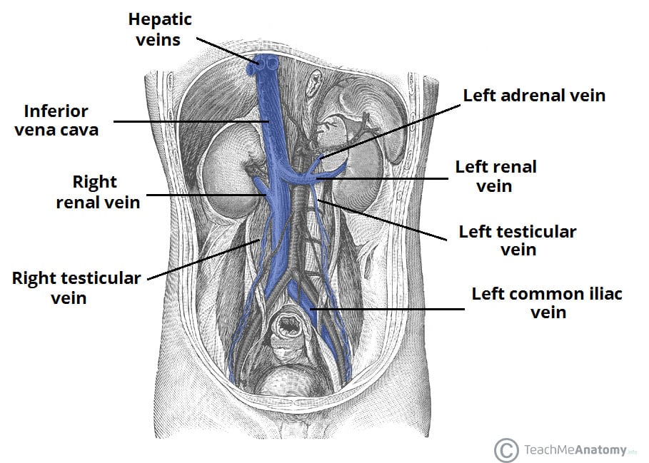
Inferior vena cava: Anatomy and function - Kenhub Feb 22, 2022 · The inferior vena cava (IVC) is the largest vein of the human body. It is located at the posterior abdominal wall on the right side of the aorta. The IVC’s function is to carry the venous blood from the lower limbs and abdominopelvic region to the heart . Transthoracic echocardiography of the inferior vena cava ... transthoracic echocardiographic findings of fat embolism are acute right heart failure: rv and inferior vena cava dilatation and echogenic material in the right atrium and ventricle and pulmonary... Inferior Vena Cava (IVC) - Geeky Medics Overview of the inferior vena cava The IVC is formed by the union of the right and left common iliac veins. It conveys systemic venous blood from the lower limbs and pelvis, the undersurface of the diaphragm and parts of the abdominal wall. The IVC does not drain blood from the gut. Course of the IVC Heart Blood Flow | Simple Anatomy Diagram, Cardiac ... Step 1 involves the superior vena cava (SVC) and inferior vena cava (IVC). They are the main blood vessels that carry the deoxygenated venous blood from the rest of the body to the right side of the heart, specifically the right atrium.
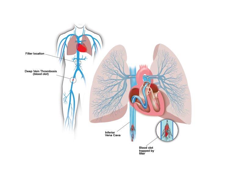
Vena cava inferior Diagram | Quizlet Start studying Vena cava inferior. Learn vocabulary, terms, and more with flashcards, games, and other study tools. Blood vessels of abdomen and pelvis : Anatomy ... - Kenhub The inferior vena cava (IVC) is the headmaster of the veins department. It collects all the blood from the abdomen, pelvis and lower limbs and carries it to the right atrium of the heart . The IVC is formed by merging of the left and right common iliac veins at the L5 vertebral level, just in front of the aortic bifurcation. Question 12 diagram - Label the following: pulmonary ... Transcribed image text: Question 12 diagram - Label the following: pulmonary artery, pulmonary vein, Superior and inferior vena cava, aorta, right and left lungs, parietal and visceral pericardium, pericardial fluid, epicardium, myocardium, endocardium, AV valves (bicuspid and tricuspid), semi lunar valves (pulmonary and aortic). Draw arrows to demonstrate the direction of blood flow through ... Inferior Vena Cava (IVC) Filter Placement | Johns Hopkins ... An inferior vena cava (IVC) filter is a small device that can stop blood clots from going up into the lungs. The inferior vena cava is a large vein in the middle of your body. The device is put in during a short surgery. Veins are the blood vessels that bring oxygen-poor blood and waste products back to the heart.
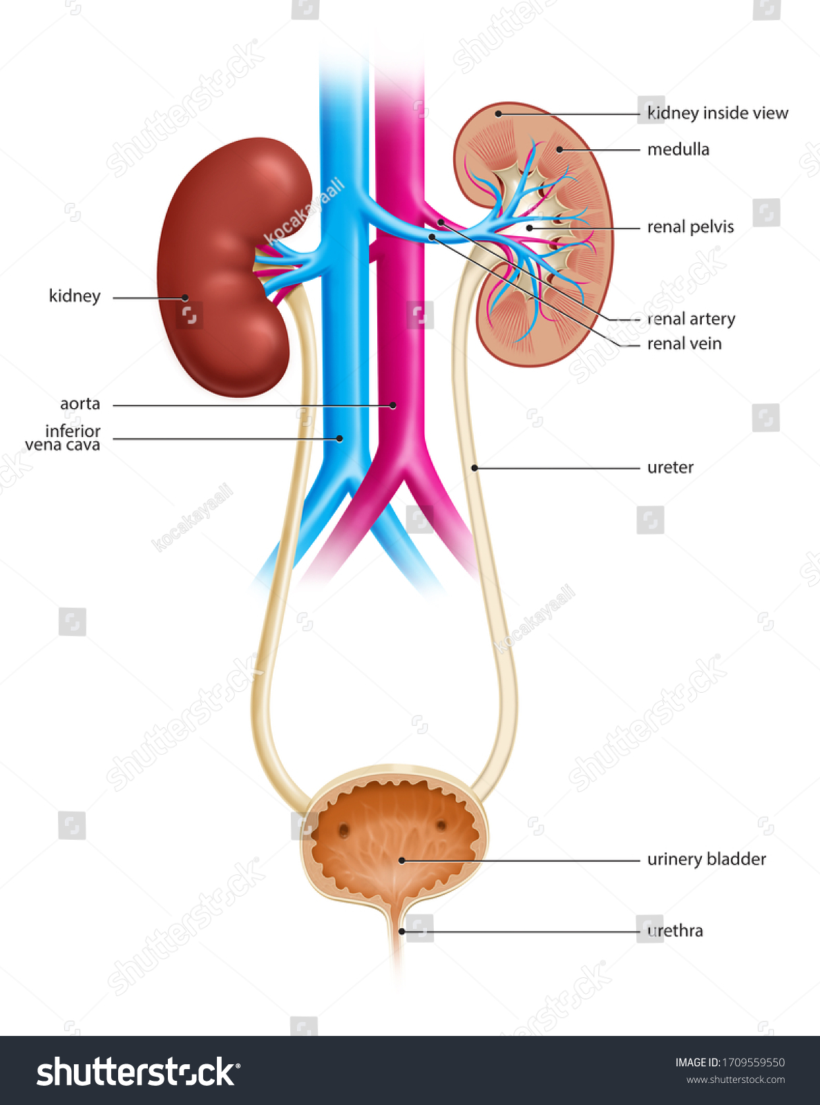
Inferior Vena Cava Function, Anatomy & Definition | Body Maps Feb 24, 2020 · The inferior vena cava runs posterior, or behind, the abdominal cavity. This vein also runs alongside the right vertebral column of the spine. The inferior vena cava is the result of two major leg...
Superior & Inferior Vena Cava Function & Location | What ... The inferior vena cava originates at around the L5 vertebrae where the left and right iliac veins converge, these vessels collect deoxygenated blood from smaller vessels below the diaphragm. From...
Flow Of Deoxygenated Blood Through The Heart - Etfatehran.net Beginning with the superior and inferior vena cavae and the coronary sinus the flowchart below summarizes the flow of blood through the heart including all arteries veins and valves that are passed along the way. Deoxygenated blood enters right atrium through Superior and Inferior Vena Cava2. The blood first enters the right atrium.
Migrated Inferior Vena Cava (IVC) Filter Strut: A Rare ... BACKGROUND Inferior vena cava (IVC) filters are indicated for patients with recurrent venous thrombosis despite proper anticoagulation or whenever anticoagulation is contraindicated. IVC filter deployment is an invasive procedure with various complications. One example is IVC filter limb fracture an …
Hepatic Veins Anatomy, Function & Diagram | Body Maps The hepatic veins carry oxygen-depleted blood from the liver to the inferior vena cava. They also transport blood that has been drained from the colon, pancreas, small intestine, and the stomach ...
From the diagram _________ is the inferior vena cava. 6 3 4 1 Solution for From the diagram _____ is the inferior vena cava. 6 3 4 1. close. Start your trial now! First week only $4.99! arrow_forward. learn. write. tutor. study resourcesexpand_more. Study Resources. We've got the study and writing resources you need for your assignments. Start exploring! ...
Heart Anatomy: Labeled Diagram, Structures, Blood Flow ... The inferior vena cava is located inferiorly, and it carries deoxygenated blood from the lower body to the right atrium. Image: Anatomy of the heart labeled diagram showing the main cardiac structures including the superior and inferior vena cava. Pulmonary Artery
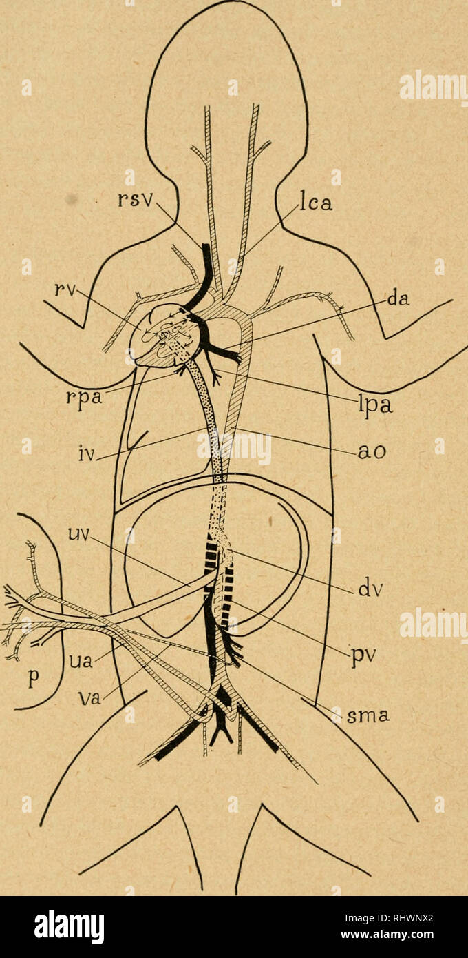
Fetal Circulation Diagram | Fetal Blood Flow & Circulatory ... The inferior vena cava takes blood to the right atrium of the heart Through a series of shunts and openings, the blood flows through the heart bypassing the lungs.
Label the Heart - Labelled diagram Label the Heart - Labelled diagram. Superior Vena Cava, Aorta, Inferior Vena Cava, Pulmonary Vein, Pulmonary Artery.
Schematic of inferior vena cava measurements during ... Schematic of inferior vena cava measurements during expiration (left) and inspiration (right). Short-axis measurements obtained after a 90 rotation of the transducer approximate the level of...
Inferior Vena Cava: Anatomy, Function, and Significance Apr 01, 2022 · The inferior vena cava (also known as IVC or the posterior vena cava) is a large vein that carries blood from the torso and lower body to the right side of the heart. From there the blood is pumped to the lungs to get oxygen before going to the left side of the heart to be pumped back out to the body.

:watermark(/images/watermark_only_sm.png,0,0,0):watermark(/images/logo_url_sm.png,-10,-10,0):format(jpeg)/images/anatomy_term/inferior-vena-cava-7/NomU64bkkS83EIiBUBanw_V._cava_inferior_02.png)


/GettyImages-184897753-ce6892a3766a4256a6951f22ef340269.jpg)

:background_color(FFFFFF):format(jpeg)/images/library/12506/Horseshoe_kidney.png)


:max_bytes(150000):strip_icc()/the-structure-of-the-vein-wall-87395965-58963cc63df78caebc05eee0.jpg)










0 Response to "41 inferior vena cava diagram"
Post a Comment