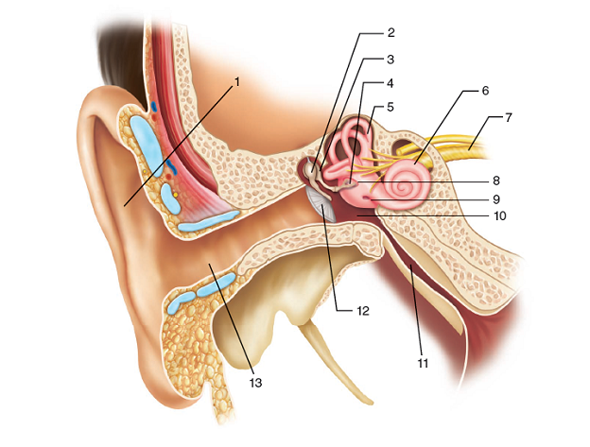
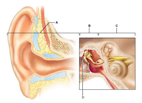
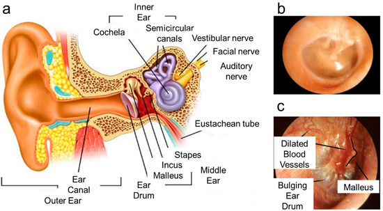
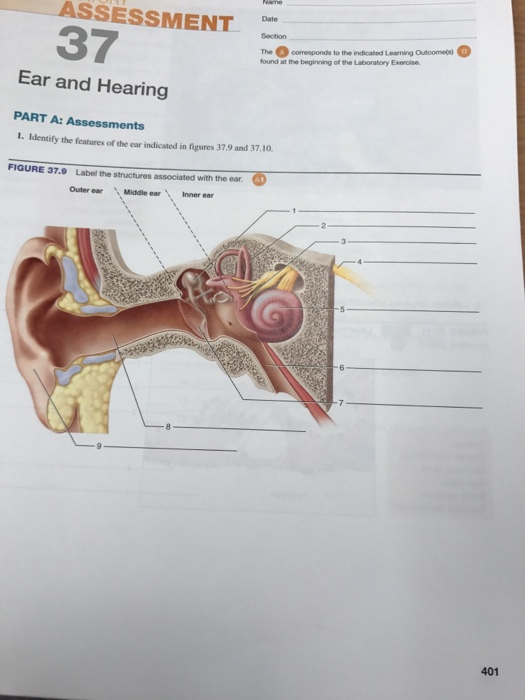
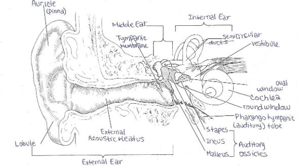
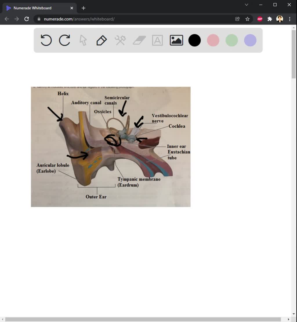
40 identify all indicated structures and ear regions in the following diagram
The Human Ear - Structure, Functions and its Parts The ear is a sensitive organ of the human body. It is mainly concerned with detecting, transmitting and transducing sound. Maintaining a sense of balance is another important function performed by the human ear. Let us have an overview of the structure and functions of the human ear. Structure of Ear. The human ear consists of three parts ... Head and neck: Regions and anatomy | Kenhub The region between these triangular regions, corresponding to the area of this broad, strap-like muscle, is the sternocleidomastoid region of the neck. The sternocleidomastoid muscle (SCM) has two heads: the rounded tendon of the sternal head which attaches to the manubrium , and the thick fleshy clavicular head which attaches to the superior ...
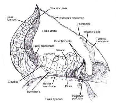
EX 25 Special Senses: hearing and Equilibrium - Quizlet Identify all indicated structures and ear regions in the following diagram refer to diagram Sacs found within the vestibule Saccule, utricle contains the spiral organ Cochlear duct Sites of the maculae Saccule, utricle Positioned in all spatial planes Semicircular ducts Hair cells of spiral organ rest on this membrane Basilar membrane

Identify all indicated structures and ear regions in the following diagram
A&P2 Lab 12 HW Flashcards - Quizlet Which of the following is the direction in which most bile would flow between the indicated points? Most bile would flow from C to A for storage and from A to B for secretion Drag the labels onto the diagram to identify the anatomical features of the liver. Ch.25 Special senses: hearing and equilibrium - Quizlet STRUCTURE COMPOSING THE EXTERNAL EAR PINNA (AURICLE), EXTERNAL AUDITORY CANAL, TYMPANIC MEMBRANE STRUCTURES COMPOSING THE INTERNAL EAR COCHLEA, SEMICIRCULAR CANALS, VESTIBULE COLLECTIVELY CALLED THE OSSICLES INCUS (ANVIL), MALLEUS (HAMMER), STAPES (STIRRUP) INVOLVED IN EQUALIZING THE PRESSURE IN THE MIDDLE EAR WITH ATMOSPHERIC PRESSURE [Solved] 386 Review Sheet 25 2. Identify the structures of ... A. Middle ear - it mainly consists of 3 bones, which help in amplification and transmission of sound wave from tympanic membrane to cochlea. The 3 bones are - malleus, incus and stapes B. Inner ear It consists of cochlea - it contains receptor for hearting. Semicircular canals and vestibule - help in balance. Step-by-step explanation
Identify all indicated structures and ear regions in the following diagram. fluidcontainedwithinthebonylabyrinthandbathingthe ... 4. Identify all indicated structures and ear regions in the following diagram. 5. Match the membranous labyrinth structures listed in column B with the descriptive statements in column A. Some terms are used more than once. Human Ear Anatomy - Parts of Ear Structure, Diagram and ... The external (outer) ear consists of the auricle, external auditory canal, and eardrum (Figure 1 and 2). The auricle or pinna is a flap of elastic cartilage shaped like the flared end of a trumpet and covered by skin. The rim of the auricle is the helix; the inferior portion is the lobule. Ligaments and muscles attach the auricle to the head. Chapter 15-The Special Senses Diagram | Quizlet Terms in this set (52) *Diagram -- *Diagram -- Identify the muscle that is controlled by the abducens nerve (CN VI). (Image #1) A B C D B (Lateral rectus muscle) Identify the muscle responsible for depressing the eye and turning it laterally. (Image #1) A B C D A (Superior oblique muscle) Name the muscle at D. (Image #1) lateral rectus Diagram of the Brain and its Functions - Bodytomy All cranial nerves are situated in the brain stem. The hindbrain is made up of the brain stem and the cerebellum. Pons. The word 'pons' literally means bridge. This is the structure that helps connect the two parts of the medulla oblongata and is often seen as a slight bulge present just above the medulla oblongata.
Ear Anatomy: Pictures Flashcards - Quizlet Identify I: Semicircular Canals. Identify J: pinna/auricle. Identify A: Cochlea. Identify I: a coiled, bony, fluid-filled tube in the inner ear through which sound waves trigger nerve impulses. External Auditory meatus. 6.3 Bone Structure - Anatomy & Physiology Figure 6.3.3 - Anatomy of a Flat Bone: This cross-section of a flat bone shows the spongy bone (diploë) covered on either side by a layer of compact bone. Osseous Tissue: Bone Matrix and Cells Bone Matrix Osseous tissue is a connective tissue and like all connective tissues contains relatively few cells and large amounts of extracellular matrix. Structure and Functions of the Ear Explicated With ... Outer ear is divided into the pinna and the external auditory meatus. The pinna, also known as the auricle is the external ear part that is located and seen on each side of our head. It is made up of cartilage and soft tissue. This helps in maintaining a particular ear shape and remains pliable. Using the above referenced figures of the anatomy of a ... Using the above-referenced figures of the anterior (left side) and posterior (right side) views of the axial skeleton, identify the specified labeled part indicated in each of the following questions. 15) Identify the structure indicated by Label E. Using the above-referenced diagram depicting the anatomical distribution of the parasympathetic ...
PDF 2 the Anatomy and Physiology of The Ear and Hearing The ear canal is about 4 centimetres long and consists of an outer and inner part. The outer portion is lined with hairy skin containing sweat glands and oily sebaceous glands which together form ear wax. Hairs grow in the outer part of the ear canal and they and the wax serve as a protective barrier and a disinfectant. 1.4 Anatomical Terminology - Anatomy & Physiology Figure 1.4.1 - Regions of the Human Body: The human body is shown in anatomical position in an (a) anterior view and a (b) posterior view. The regions of the body are labeled in boldface. A body that is lying down is described as either prone or supine. Human Ear: Structure and Functions (With Diagram) ADVERTISEMENTS: In this article we will discuss about the structure and functions of human ear. Structure of Ear: Each ear consists of three portions: (i) External ear, ADVERTISEMENTS: (ii) Middle ear and (iii) Internal ear. 1. External Ear: It comprises a pinna, external auditory meatus (canal) & tympanic membrane. (i) Pinna: ADVERTISEMENTS: The pinna is […] Practical worksheet 5 (JAN 2021) The Sensory System ... Identify all indicated structures and ear regions that are provided with leader lines or brackets in the following diagram. 5. Match the membranous labyrinth structures in the listed key with the descriptive statements below.
[Solved] 386 Review Sheet 25 2. Identify the structures of ... A. Middle ear - it mainly consists of 3 bones, which help in amplification and transmission of sound wave from tympanic membrane to cochlea. The 3 bones are - malleus, incus and stapes B. Inner ear It consists of cochlea - it contains receptor for hearting. Semicircular canals and vestibule - help in balance. Step-by-step explanation
Ch.25 Special senses: hearing and equilibrium - Quizlet STRUCTURE COMPOSING THE EXTERNAL EAR PINNA (AURICLE), EXTERNAL AUDITORY CANAL, TYMPANIC MEMBRANE STRUCTURES COMPOSING THE INTERNAL EAR COCHLEA, SEMICIRCULAR CANALS, VESTIBULE COLLECTIVELY CALLED THE OSSICLES INCUS (ANVIL), MALLEUS (HAMMER), STAPES (STIRRUP) INVOLVED IN EQUALIZING THE PRESSURE IN THE MIDDLE EAR WITH ATMOSPHERIC PRESSURE
A&P2 Lab 12 HW Flashcards - Quizlet Which of the following is the direction in which most bile would flow between the indicated points? Most bile would flow from C to A for storage and from A to B for secretion Drag the labels onto the diagram to identify the anatomical features of the liver.











:watermark(/images/watermark_only_sm.png,0,0,0):watermark(/images/logo_url_sm.png,-10,-10,0):format(jpeg)/images/anatomy_term/meatus-acusticus-externus-2/cNYVakPkJkzvw8J3mO7HA_Meatus_acusticus_externus_01.png)





0 Response to "40 identify all indicated structures and ear regions in the following diagram"
Post a Comment