41 gram negative cell wall diagram
Solved In figure 4.3 which diagram of a cell is a gram ... Thus, the answer is c. b Explaination: 1] In Gram negative cell wall a composed …. View the full answer. Transcribed image text: Environment b. Plasma membrane inside cell in Figure 4.3, which diagram of a cell wall is a gram-negative ceil wall? Select one 口a, neither a nor b b The answer cannot be determined based on the informa tan provided. Differences between Gram Positive and Gram Negative Bacteria Gram Negative. 1. Gram Reaction. Retain crystal violet dye and stain blue or purple. Can be decolorized to accept counterstain (safranin) and stain pink or red. 2. Cell Wall. Cell Wall is 20-30 nm thick. Cell Wall is 8-12 nm thick.
Diagram demonstrating of the cell wall structure of (a ... Download scientific diagram | Diagram demonstrating of the cell wall structure of (a) Grampositive bacterium, (b) Gram-negative bacterium, and (c) mycobacterium. from publication: Differentiation ...

Gram negative cell wall diagram
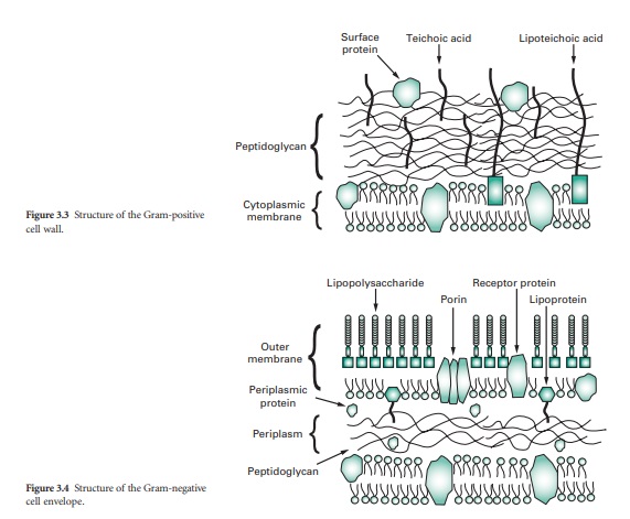
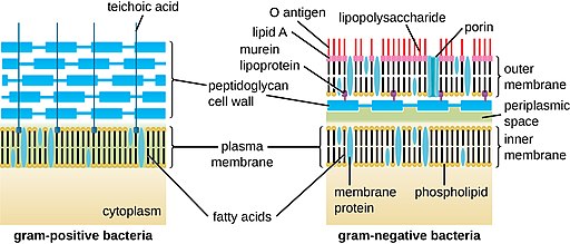
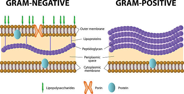
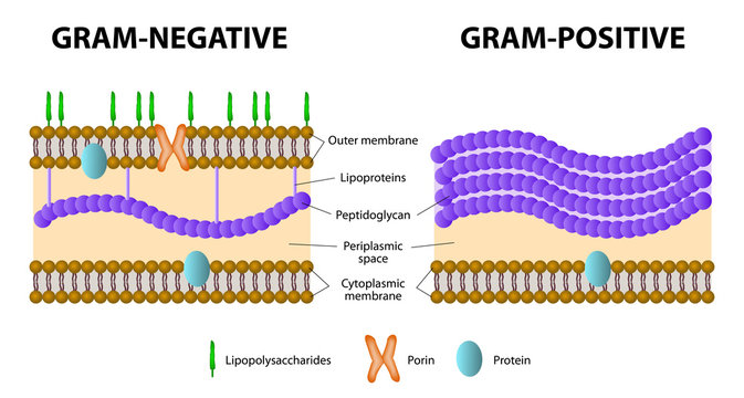
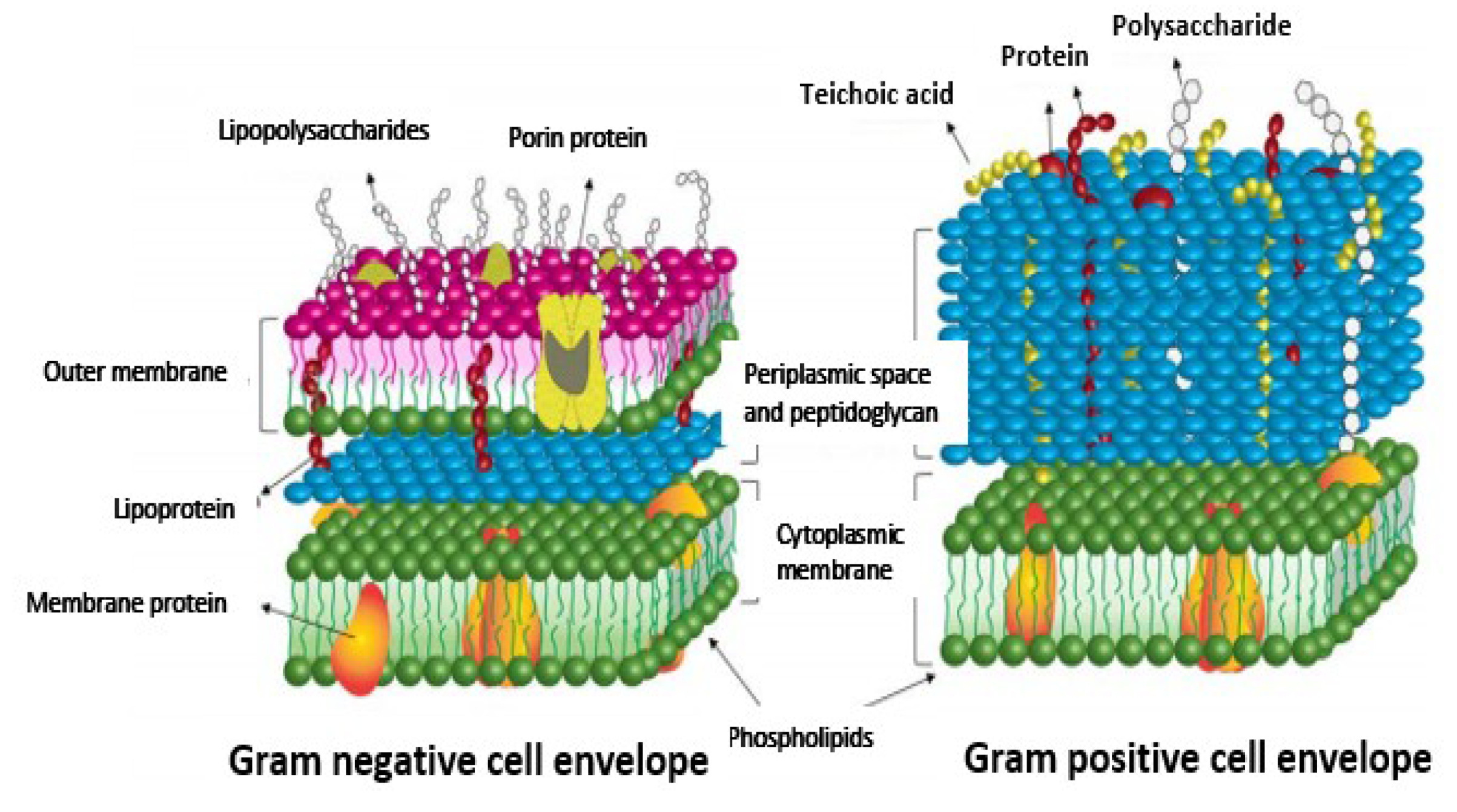
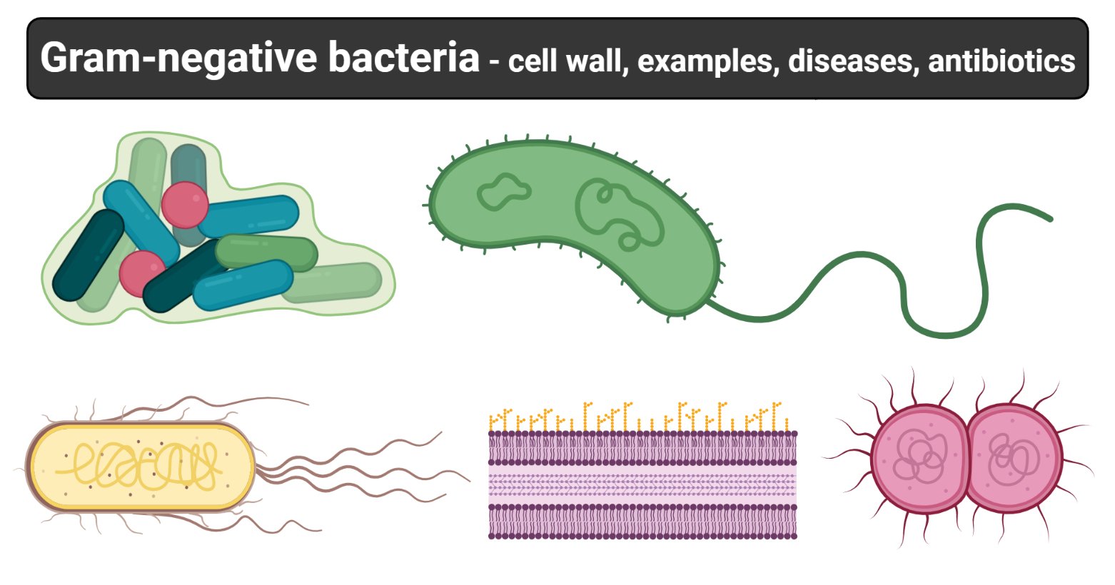
Gram negative bacteria examples, list, and structure ... Gram negative bacteria include some bacterial cell examples that possess a thin peptidoglycan cell wall structure. They can cause diseases in humans and stain pink in a gram stain test. In other words, gram-negative bacteria can be defined as the bacterial cells that do not take up the crystal violet stain used in the Gram staining procedure and therefore stain pink at the end of the procedure. Difference Between Gram Positive and Gram Negative Cell Wall The key difference between gram positive and gram negative cell wall is that the gram positive cell wall has a thick peptidoglycan layer with teichoic acids while gram negative cell wall has a thin peptidoglycan layer surrounded by an outer membrane. Another major difference between gram positive and gram negative cell wall is that the gram positive cell wall stains in purple colour in grams ... Microbiology Ch. 4 Flashcards & Practice Test - Quizlet In Figure 4.3, which diagram of a cell wall is a gram-negative cell wall? B. In Figure 4.3, which diagram of a cell wall possesses lipid A/endotoxin responsible for symptoms associated with infection. B. In Figure 4.3, which diagram of a cell wall has a structure that protects against osmotic lysis?
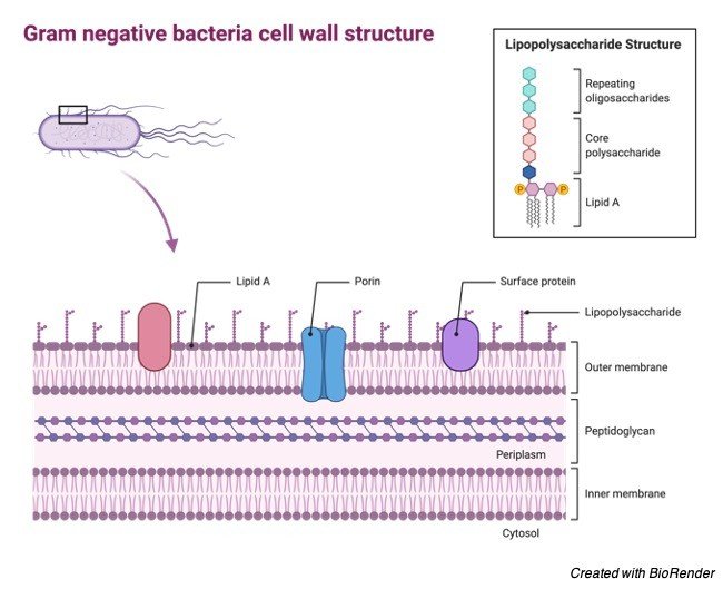
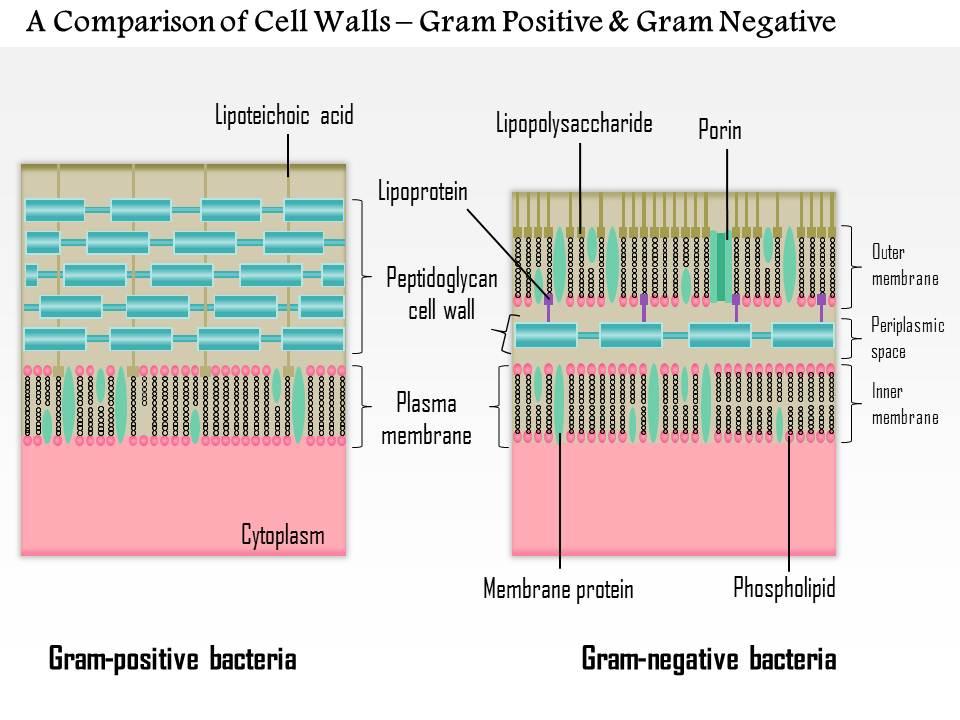
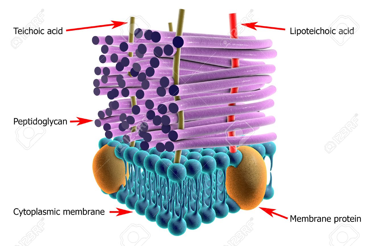
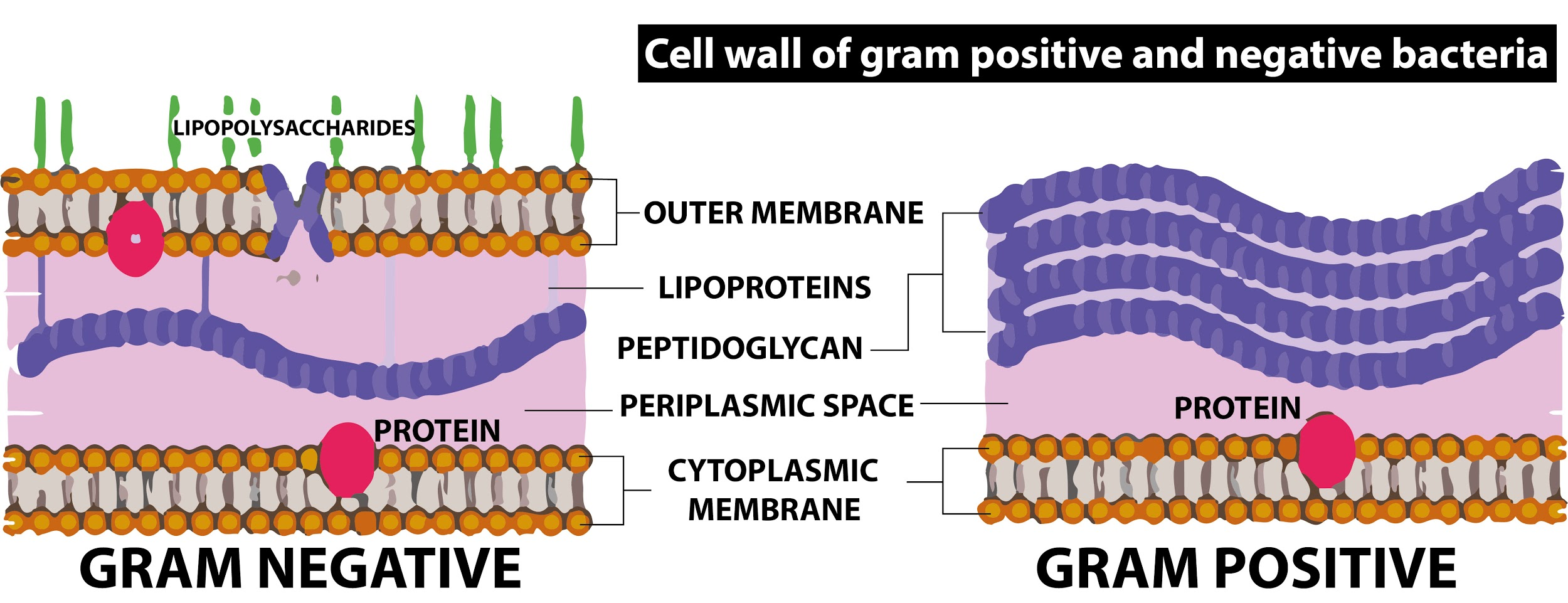
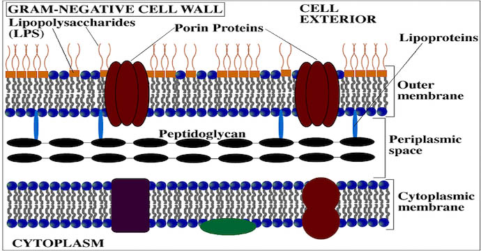
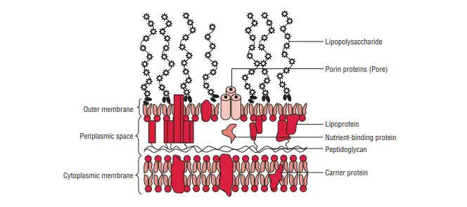
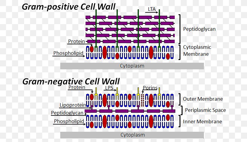
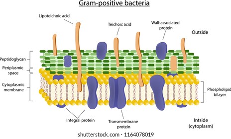
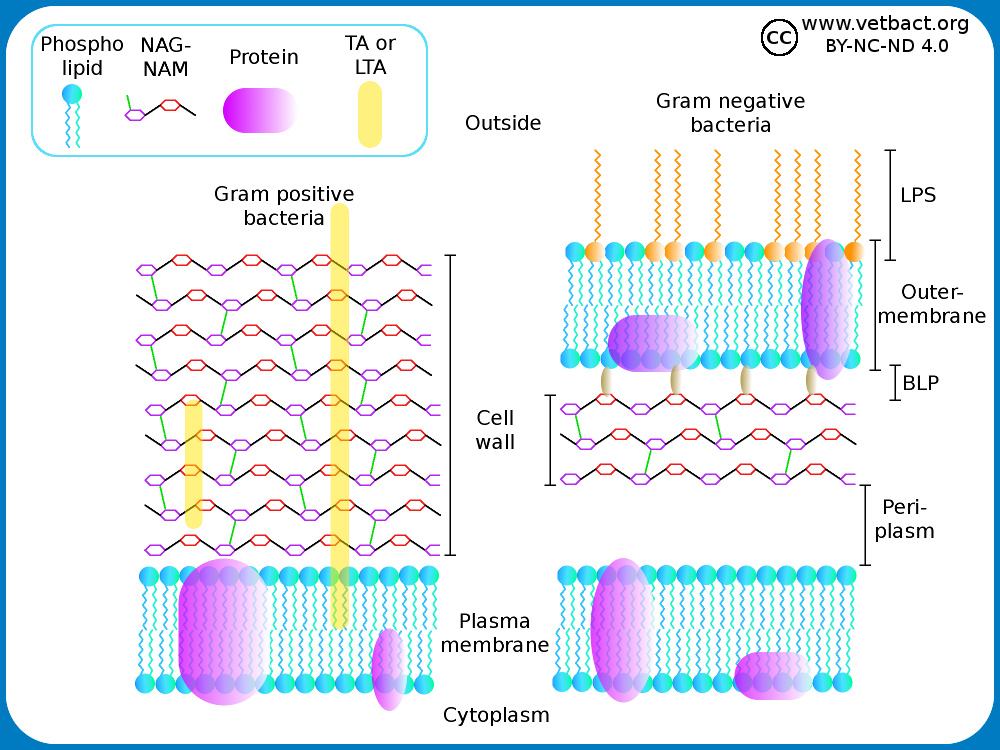
Gram negative cell wall diagram. Bacteria shapes, structure and diagram - Jotscroll The gram-Positive Cell wall of Bacteria. Bacterial cell wall that is gram-positive contains peptidoglycan and teichoic acids with some species having additional carbohydrates and proteins. The murein component is what gives shape to the gram-positive bacterial cell wall; it also helps the bacteria cells to resist osmotic pressure. Major Difference Between Gram-Positive and Gram-Negative ... In gram-negative bacteria, the cell wall is made up of an outer membrane and several layers of peptidoglycan. The outer membrane is composed of lipoproteins, phospholipids, and LPS. The peptidoglycan stays intact to lipoproteins of the outer membrane that is located in the fluid-like periplasm between the plasma membrane and the outer membrane. Structure of Gram-negative cell wall - microbiologynote.com Feb 13, 2022 · Schematic diagram of the cell wall of the Gram-negative bacteria. Outer membrane The membrane’s outer layer has a two-layered design. its inner layer is similar the composition of the membrane of cells, and the outer membrane is made up of a distinct component known as lipopolysaccharide. Bacterial Cell Wall Structure: Gram-positive & negative In Gram-positive bacteria, peptidoglycan makes up as much as 90% of the thick cell wall enclosing the plasma membrane. Gram Staining! During Gram staining, these thick, multiple layers (20-80 nm) of peptidoglycan retain the dark purple primary stain crystal violet, whereas Gram-negative bacteria stain pink.
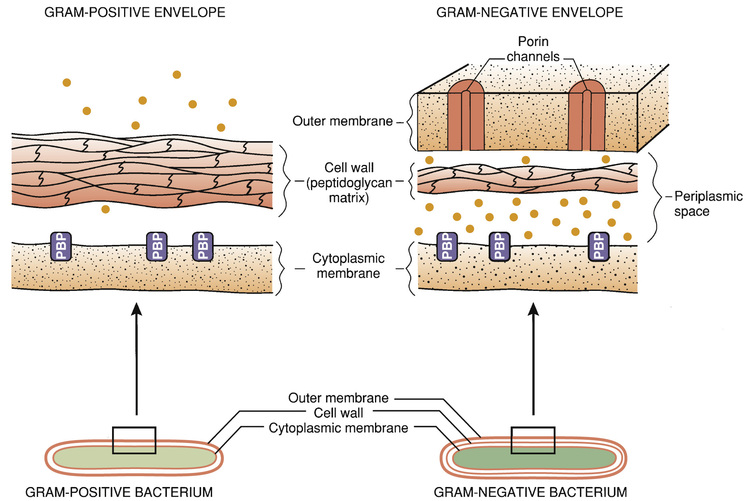
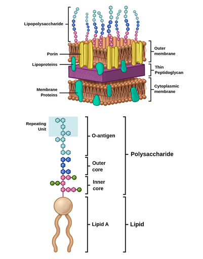
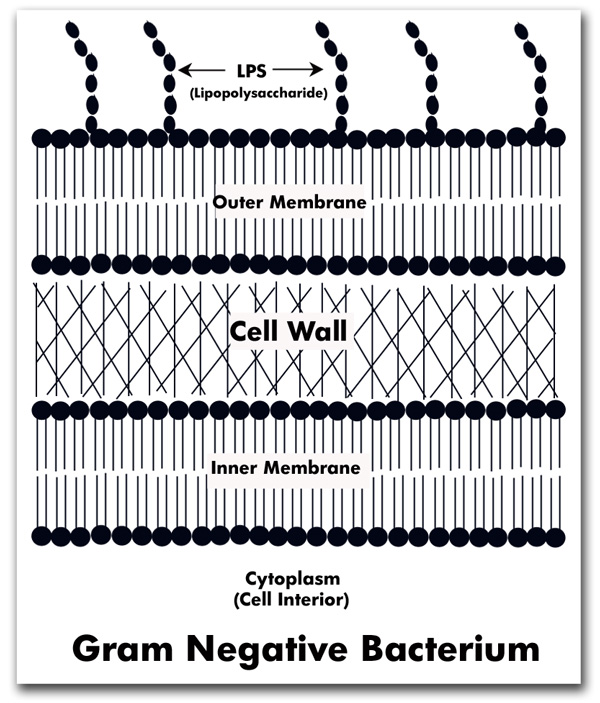
Gram Positive vs. Gram Negative Bacteria - ThoughtCo Gram positive bacteria have cell walls composed of thick layers of peptidoglycan. Gram positive cells stain purple when subjected to a Gram stain procedure. Gram negative bacteria have cell walls with a thin layer of peptidoglycan. The cell wall also includes an outer membrane with lipopolysaccharide (LPS) molecules attached. Gram Positive vs Gram Negative | Technology Networks Gram negative bacteria appear a pale reddish color when observed under a light microscope following Gram staining. This is because the structure of their cell wall is unable to retain the crystal violet stain so are colored only by the safranin counterstain. Examples of Gram negative bacteria include enterococci, salmonella species and ... Difference between Gram Positive and Gram Negative Bacteria In gram-negative bacteria, the cell wall is consisting of an outer membrane and several layers of peptidoglycan. Whereas the outer membrane is composed of lipoproteins and phospholipids. The peptidoglycan stays intact to the lipoproteins of the outer membrane. It is located in the fluid-like periplasm between the plasma membrane and the outer ... Bacterial Cell Walls Function & Parts | What is a ... Gram-negative bacteria - stain pink with the Gram stain, are double membraned (outer membrane present), have a thin peptidoglycan layer, lipopolysaccharide (LPS) in the cell wall, with a periplasm ...
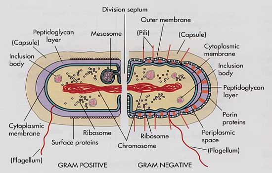
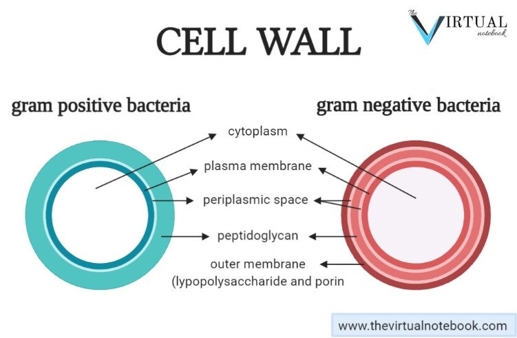
File:Gram negative cell wall.svg - Wikipedia This file is licensed under the Creative Commons Attribution-Share Alike 4.0 International, 3.0 Unported, 2.5 Generic, 2.0 Generic and 1.0 Generic license.: You are free: to share - to copy, distribute and transmit the work; to remix - to adapt the work; Under the following conditions: attribution - You must give appropriate credit, provide a link to the license, and indicate if changes ... Structures of Gram-Negative Cell Walls and Their Derived ... Gram-negative cell walls are strong enough to withstand ∼3 atm of turgor pressure (), tough enough to endure extreme temperatures and pHs (e.g., Thiobacillus ferrooxidans grows at a pH of ≈1.5) and elastic enough to be capable of expanding several times their normal surface area ().Strong, tough, and elastic … the gram-negative cell wall is a remarkable structure which protects the ... Gram positive versus negative cell wall structure ... Gram positive versus negative cell wall structure differences outline diagram. 1. Editable Vector .AI file. 2. Editable Vector .EPS-10 file. 3. High-resolution JPG image. Use for everything except reselling item itself. Description: Gram positive versus negative cell wall structure differences outline diagram. Bacteria: Cell Walls - General Microbiology 4 Bacteria: Cell Walls . It is important to note that not all bacteria have a cell wall.Having said that though, it is also important to note that most bacteria (about 90%) have a cell wall and they typically have one of two types: a gram positive cell wall or a gram negative cell wall.. The two different cell wall types can be identified in the lab by a differential stain known as the Gram stain.
Bacterial Cell Envelope
Gram Stain Technique (Theory) : Microbiology Virtual Lab I ... Gram Negative Cell Wall: Gram-negative bacteria have a thinner layer of peptidoglycan (10% of the cell wall) and lose the crystal violet-iodine complex during decolorization with the alcohol rinse, but retain the counter stain Safranin, thus appearing reddish or pink. They also have an additional outer membrane which contains lipids, which is ...
Cell Wall - Bacteria
Cell Wall of Bacteria: Structure, Functions, Gram Positive ... The walls of gram-positive bacteria have simpler chemical structures compared to gram-negative bacteria. Gram-positive cell wall. Gram-positive cell wall is thick measuring about 15-80 nm and more homogenous compared to gram-negative cell wall. This cell wall consists of large amount of peptidoglycan arranged in several layers. Peptidoglycan in ...
Cell Wall - Gram Positive vs Negative Bacteria | Easy Biology ...
MICROBIOLOGY CH. 4 Flashcards - Quizlet In Figure 4.3, which diagram of a cell wall is a gram-negative cell wall? A) a B) b C) both a and b D) neither a nor b E) The answer cannot be determined based on the information provided. B) b. In Figure 4.3, which diagram of a cell wall possesses lipid A/endotoxin responsible for symptoms associated with infection?

Solved er 4 Multiple-Choice Question 27 Part A Environment ...
Do Gram Negative Bacteria Have Thick Cell Walls ... Do Gram-Positive Bacteria Have Thinner Cell Walls? Gram negative and Gram positive bacteria have different structures in the diagram below. In Gram positive bacteria, a peptidoglycan layer is thick, and not an lipid membrane is present on the outer layer. Gram negative bacteria have both a peptidoglycan layer and a lipids layer on their surfaces.

Gram Staining | BioNinja
Gram Positive Vs Gram Negative Bacteria: A Comparison ... Ø Both groups possess capsule. Ø In both groups, cell wall is made up of peptidoglycan Learn more: Peptidoglycan vs Pseudo-peptidoglycan Ø In both groups, cytoplasm is surrounded by lipid bilayer with many membrane spanning proteins. Ø Both gram-positive and gram-negative bacteria commonly have a surface layer called an S-layer. Ø Both groups of bacteria undergo genetic recombination ...
Bacterial Cell Wall Structure: Gram Positive vs Gram Negative
PDF Bacterial Cell Structure - Bellarmine University 3.4 Bacterial Cell Walls 1. Describe peptidoglycan structure. 2. Compare and contrast the cell walls of typical Gram-positive and Gram-negative bacteria. 3. Relate bacterial cell wall structure to the Gram-staining reaction. 37
0614 A Comparison Of The Cell Walls Gram Positive And Gram ...
Solved Complete this Venn diagram comparing Gram Positive ... 100% (2 ratings) Answer: Follow the labels marked in the below diagram to understand the points mentioned in the table. A: Gram Positive Bacteria Cell Wall B: Similarities C: Gram Negative Bacteria Cell Wall A B C Teichoic acid is present in the peptidoglycan layer P …. View the full answer.
Bacterial Structure - WikiEducator
Schematic structure of Gram-positive and Gram-negative ... Download scientific diagram | Schematic structure of Gram-positive and Gram-negative cell walls. Gram-positive cell walls contain only one lipid plasma membrane and a thick peptidoglycan layer ...
Drugs That Weaken the Bacterial Cell Wall I | Basicmedical Key
Gram-positive Cell Wall Vs Gram-negative Cell Wall ... Gram staining Procedure. Retains the crystal violet dye and appear purple in colour. Does not retain the crystal violet dye and appear pink in colour. These differences between the Cell Wall of Gram-positive and Gram-negative Bacteria are classified based on their structure, composition of the cell and by the procedure of Gram staining technique.

Pin em MediMoon.com
Gram Positive vs. Negative Bacteria | Overview ... In regards to Gram positive vs Gram negative cell wall, Gram positive bacteria have a thick cell wall made of peptidoglycan. The Gram positive cell wall is a rigid structure, located just outside ...
Structure Of Cell Wall Of Gram-positive Bacteria, 3D ...
Gram-negative bacteria- cell wall, examples, diseases ... Apr 11, 2021 · The cell wall of gram-negative bacteria is complex having a thin layer of the peptidoglycan layer of 2-7nm and a thick outer membrane of 7-8nm thick. Microscopically, there is a space that is seen between the cell membrane and the cell wall, known as the periplasmic space made up of periplasm.

Microbiological educational diagram sample: Cell envelope of ...
2.3B: The Gram-Negative Cell Wall - Biology LibreTexts Jan 03, 2021 · In electron micrographs, the Gram-negative cell wall (Figures 1) appears multilayered. It consists of a thin, inner wall composed of peptidoglycan and an outer membrane. Figure 2.3 B. 1 (left): Electron Micrograph of a Gram-Negative Cell Wall (right) Structure of a Gram-Negative Cell Wall.

a) Diagram of a gram-negative cell wall. (b) Electron ...
Microbiology Ch. 4 Flashcards & Practice Test - Quizlet In Figure 4.3, which diagram of a cell wall is a gram-negative cell wall? B. In Figure 4.3, which diagram of a cell wall possesses lipid A/endotoxin responsible for symptoms associated with infection. B. In Figure 4.3, which diagram of a cell wall has a structure that protects against osmotic lysis?

Cell Wall Structure of Gram-negative Bacteria for Example ...
Difference Between Gram Positive and Gram Negative Cell Wall The key difference between gram positive and gram negative cell wall is that the gram positive cell wall has a thick peptidoglycan layer with teichoic acids while gram negative cell wall has a thin peptidoglycan layer surrounded by an outer membrane. Another major difference between gram positive and gram negative cell wall is that the gram positive cell wall stains in purple colour in grams ...
Give the difference between the cell walls of grampositive ...
Gram negative bacteria examples, list, and structure ... Gram negative bacteria include some bacterial cell examples that possess a thin peptidoglycan cell wall structure. They can cause diseases in humans and stain pink in a gram stain test. In other words, gram-negative bacteria can be defined as the bacterial cells that do not take up the crystal violet stain used in the Gram staining procedure and therefore stain pink at the end of the procedure.

Gram Negative Cell Wall Diagram | Quizlet

BACTERIAL CELL WALL - Microbiology Class
2,177 Gram Negative Bacteria Stock Photos, Pictures & Royalty ...
Bacterial Cell Wall Images – Browse 396 Stock Photos, Vectors ...

cell wall structure of Gram-negative Bacteria for example ...
Gram-negative cell wall - Labster Theory

File:Gram negative cell wall.svg - Wikimedia Commons

Pathogen Recognition and Innate Immunity: Cell

Gram negative bacteria examples, list, and structure - Jotscroll

Gram Positive vs. Gram Negative Bacteria | Gram negative ...

Microbiological educational diagram sample: Cell envelope of ...
What is the Difference Between Gram Positive and Gram ...
Experiment 4A | Lab04 | Virtual Edge| Molb 2021 | College of ...

File:Gram negative cell wall-nl.svg - Wikimedia Commons
How can we change gram negative to gram positive in bacteria ...

Bacteria cell wall gram positive and gram negative

Morphology of gram positive and gram negative cell wall - YouTube
Structure of Gram-negative cell wall
Molecules | Free Full-Text | Application of Porphyrins in ...
Bacterial Cell Structure Cell Wall Gram-positive Bacteria ...

Endotoxin Gram-negative Bacteria Gram-positive Bacteria ...
Cell Wall Structure Grampositive Bacteria Stock Vector ...

My Scientific Blog - Research and Articles: The Bacterial ...
VetBact
Gram negative bacteria, cell wall, diseases - The Virtual ...
Gram-negative bacteria- cell wall, examples, diseases ...
0 Response to "41 gram negative cell wall diagram"
Post a Comment