39 meninges of the brain diagram
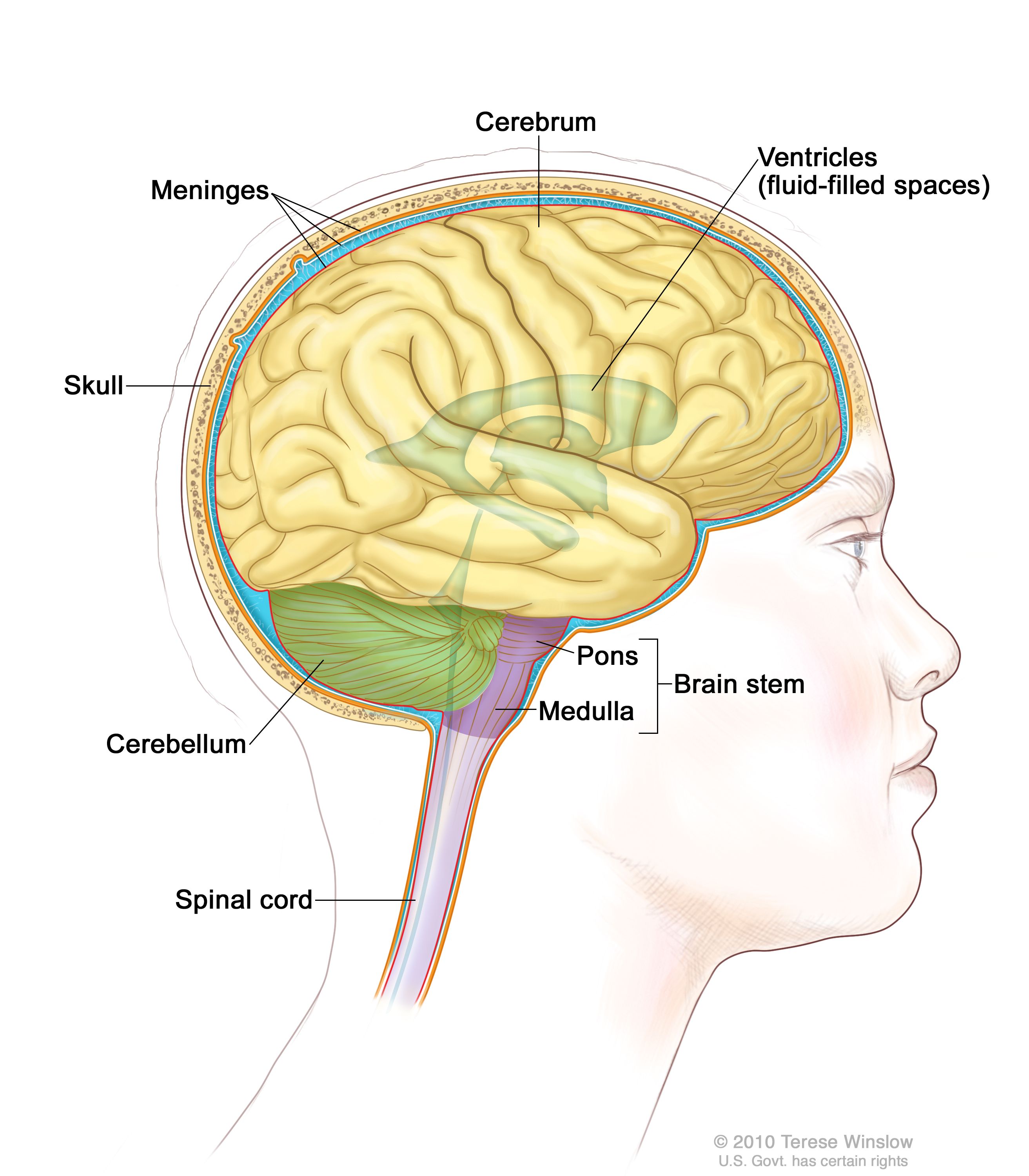
Discover how your brain controls your body and houses your mind. This allows your brain to monitor and regulate unconscious body processes, such as digestion and breathing and to coordinate most voluntary movements of your body. The cerebrum is the largest part of your brain. It sits on top of the rest of your brain, rather like a mushroom cap covering its stalk. Meninges - an overview | ScienceDirect Topics Meninges-brain interface: signals from the meninges regulate development of the CNS 1041. Also, depending on where the expanding mass causing the increased pressure is located, certain portions of the brain may herniate from one side of a dural reflection to another (Figs.
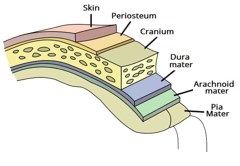
IX. Neurology. 4g. The Meninges of the Brain and Medulla Spinalis. The Cranial Dura Mater (dura mater encephali; dura of the brain) lines the interior of the skull, and serves the twofold purpose of an internal periosteum to the It is composed of two layers, an inner or meningeal and an outer or endosteal, closely connected together, except in certain situations, where...

Meninges of the brain diagram
CT Brain Anatomy - Meninges Tutorial on the anatomical location of the meninges relating to acute CT brain pathology. The falx cerebri and the tentorium cerebelli are thick infoldings of the meninges which are visible on CT imaging. Elsewhere the meningeal layers are not visible on CT as they are closely applied to the... Innervation of the meninges | Radiology Reference... | Radiopaedia.org The sensory innervation of the meninges is primarily by meningeal branches of both the trigeminal and vagus nerves with a smaller contribution from the upper cervical spinal nerves 1,2. The supratentorial dura mater is mainly supplied by the ophthalmic division of the trigeminal nerve 3. Like... Brain Coverings: Meninges | Johns Hopkins Medicine The cerebellum ("little brain") is a fist-sized portion of the brain located at the back of the head, below the temporal and occipital lobes and above the brainstem. Three layers of protective covering called meninges surround the brain and the spinal cord. The outermost layer, the dura mater, is thick and...
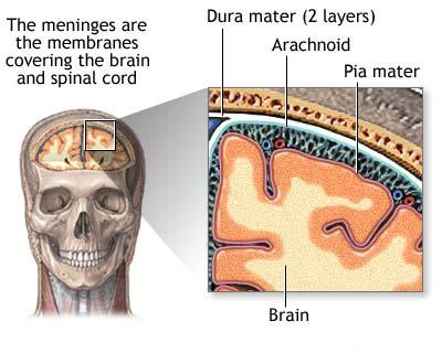
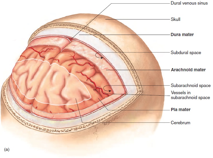
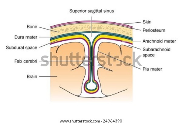
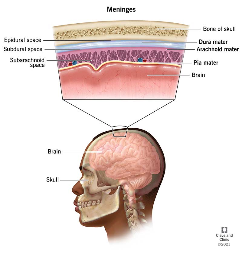
Meninges of the brain diagram. Human Brain: facts and information The brain is extremely sensitive and delicate, and so it requires maximum protection, which is provided by the hard bone of the skull and three tough membranes called meninges. The spaces between these membranes are filled with fluid that cushions the brain and keeps it from being damaged by contact... Meninges | Concise Medical Knowledge The brain and the spinal cord are enveloped by 3 overlapping layers of connective tissue called the meninges. of the meninges, and the etiology can be elicited by examining CSF, which is Image: "Diagram of section of top of brainBrainThe part of central nervous system that is contained within the... Meninges - Brain The brain is surrounded by mesodermal coverings, the meninges. The dura mater lines the inner surface of the skull and also forms the periosteum. Sturdy septa extend from it deep into the cranial cavity. Meninges and Cerebrospinal Fluid (Gross Anatomy of the Brain) Part 1 The Meninges The tissues comprising the brain and spinal cord are very delicate and require special protection. This is provided by the bony cranial Sheet-like processes, called septa, extend from the meningeal layer of the dura deep into the cranial cavity, forming freely communicating compartments.
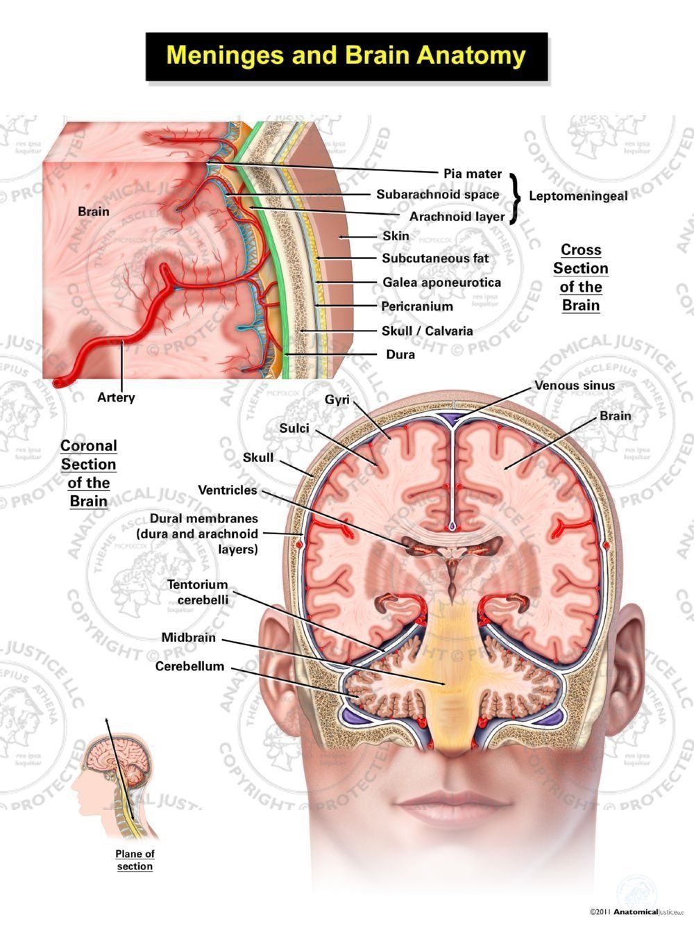
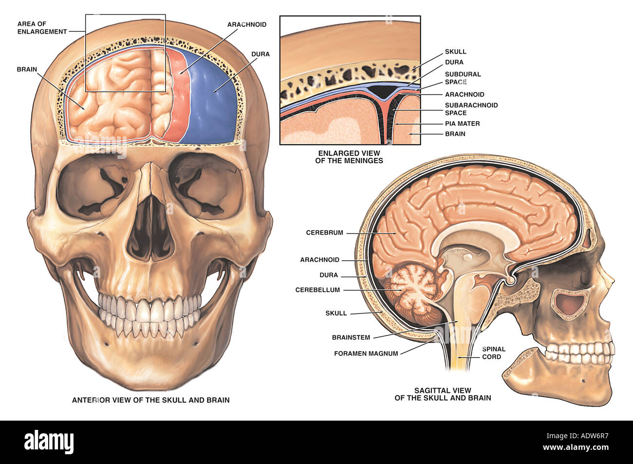
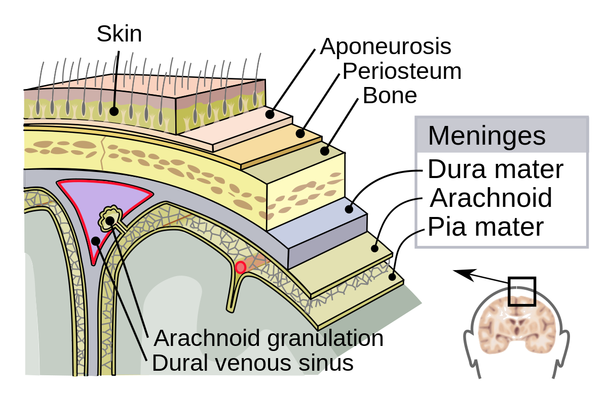
The Meninges - Dura - Arachnoid - Pia - TeachMeAnatomy The meninges refer to the membranous coverings of the brain and spinal cord, comprised of the dura, arachnoid and pia mater. The meningeal layer of dura mater folds inwards upon itself to form four dural reflections. These reflections project into the cranial cavity, dividing it into several compartments... Brain - Human Brain Diagrams and Detailed Information Our interactive diagram helps you explore the anatomy of the human brain and learn all about how it functions. Three layers of tissue, collectively known as the meninges, surround and protect the brain and spinal cord. The dura mater forms the leathery, outermost layer of the meninges. Membranes of The Brain (Meninges) - Earth's Lab The brain and spinal cord are enclosed inside 3 protective membranes referred to as meninges. From without inward these are as follows: Dura mater, Arachnoid mater and Pia mater. Originally, the... Meninges notes and explanations - MENINGES The brain... - StuDocu The brain is surrounded by three meninges (meninges encephali): the outer - the dura mater, the. middle - arachnoid (arachnoid) and the inner, adjacent subarachnoid spaces of the brain and spinal cord. Cerebrospinal fluid is formed in the vessels as a result of water seeping and certain components...
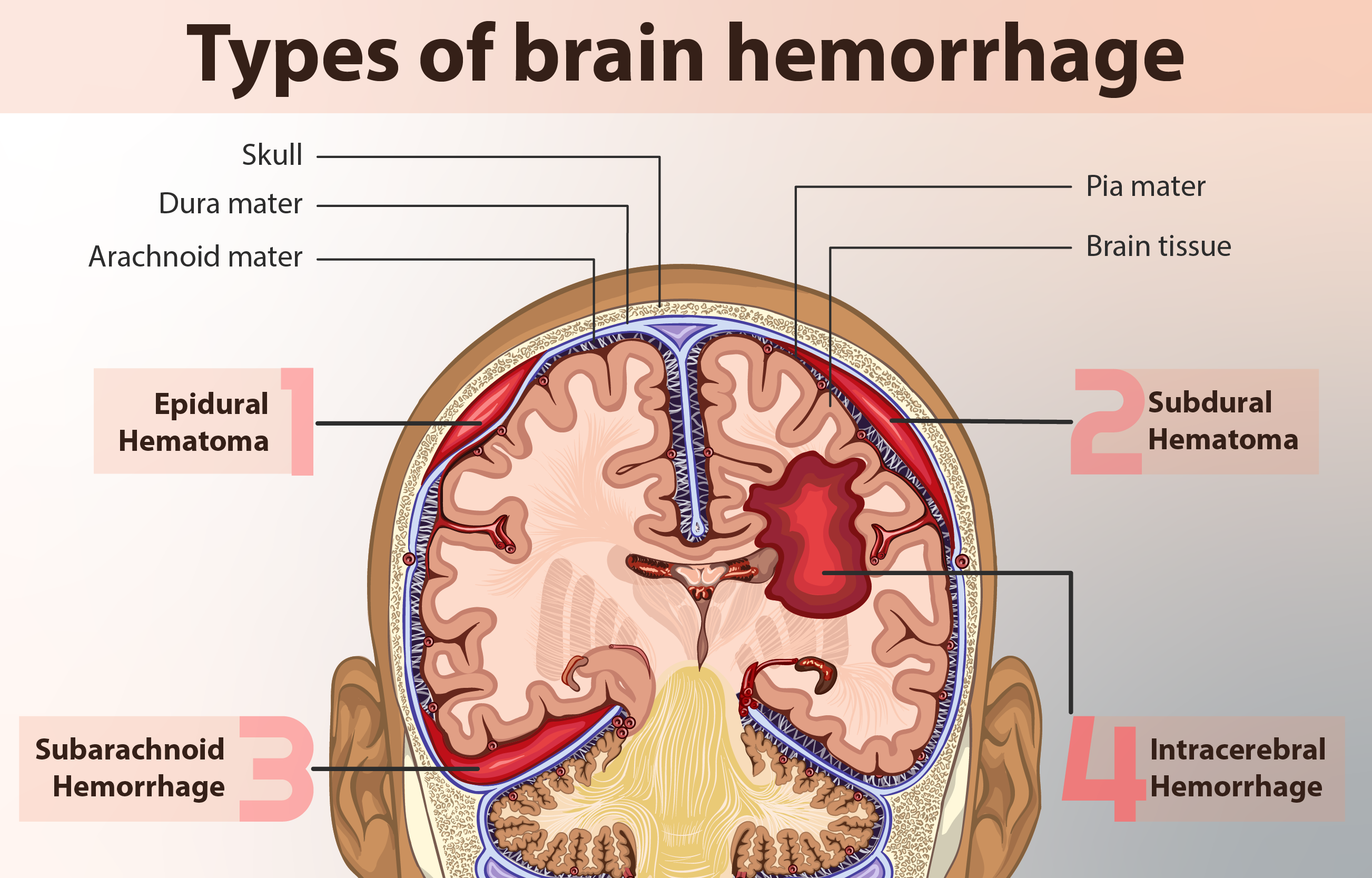
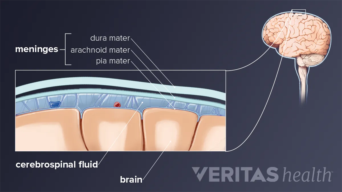
Meninges - Wikipedia In anatomy, the meninges are the three membranes that envelop the brain and spinal cord. In mammals, the meninges are the dura mater, the arachnoid mater, and the pia mater. Cerebrospinal fluid is located in the subarachnoid space between the arachnoid mater and the pia mater. Brain Meninges | Facts, Blood & Nerve Supply, Hemorrhages Meninges are the capsule-like coverings that provide protection to the brain and spinal cord. The meningeal layer sends inward septa that divide the cranial cavity into different chambers. These chambers lodge different parts of the brain such as the cerebellum and cerebral hemispheres. Cranium, Ventricles, and Meninges | BRAIN adheres to brain surface closely and follows gyri and depths of sulci. surrounds initial portion of each blood vessel as it enters brain surface, , then fuses Spinal cord- enveloped by same 3 meningeal layers as exits through foramen of magnum; only difference is a layer of epidural fat in the spinal canal. Brain (Human Anatomy): Picture, Function, Parts, Conditions, and More WebMD's Brain Anatomy Page provides a detailed diagram and definition of the brain including its function, parts, and conditions that affect it. The occipital lobes contain the brain's visual processing system. The brain is surrounded by a layer of tissue called the meninges.
Brain Meninges Skull Cranial cavity Anatomy, meninges of the ...
meninges | anatomy | Britannica meninges, three membranous envelopes—pia mater, arachnoid, and dura mater—that surround the brain and spinal cord. The pia mater is the meningeal envelope that firmly adheres to the surface of the brain and spinal cord. It is a very thin membrane composed of fibrous tissue covered on its outer...
File:Meninges diagram.jpg - Wikimedia Commons
Human Brain: Facts, Functions & Anatomy | Live Science A medical illustration of the human brain from 'Quain's Elements of Anatomy, Eighth Edition, Vol.II' (by William Sharpey MD, LLD, FRS L&E, Allen Thomson, MD, LLD, FRS L&E, and Edward Albert Schafer) depicts the right half of the brain, 1876. (Image credit: Vintage MedStock/Getty Images).

10-11 The Meninges Diagram | Quizlet
Meninges Of The Brain Diagram - Brain Meninges Skull Cranial... User disneyxdbernstein uploaded this Meninges Of The Brain Diagram - Brain Meninges Skull Cranial Cavity Anatomy PNG PNG image on December 30, 2018, 10:19 am. The resolution of this file is 1500x1035px and its file size is: 1.74 MB.

294 Meninges Stock Photos and Images - 123RF
Meninges of the Brain | 3D Anatomy Tutorial - YouTube In this tutorial we review the meninges of the brain, looking at the following structures:- Dura mater- Arachnoid mater- Subarachnoid space- Arachnoid...

Meninges brain Royalty Free Vector Image - VectorStock
How Human Brain Functions? Brain Functioning List with Diagram What are human brain functions in the body? To list all the functions and responsibilities of this collection of billions of neurons, you need to compose a whole book. The higher functions of the brain are to be performed by the cerebral cortex, a highly developed region of the brain.

Meninges of the brain | Dura mater, Brain models, Epidural
Brain This brain scan was taken after a pineal tumor was removed with the help of TB bacteria. The caverns that are created after the tumor has been The fourth ventricle between the brainstem and the cerebellum connects with the subarachnoid space (see meninges) and the central canal of the spinal...
The Meninges - Dura - Arachnoid - Pia - TeachMeAnatomy
Anatomical diagrams of the brain - eAnatomy Brain , Coronal section : Brain , Anatomy diagram. Numerous illustrations are available on the cerebellum, representation of cerebellar lobes Ventricles and meninges. Functional with the motor pathways and the senses and some brainstem nuclei. Cerebral veins and venous sinuses of the dura...

Advances in Meningeal Immunity: Trends in Molecular Medicine
Meninges of the Brain | An illustrated review Meninges of the Brain (Dura, Arachnoid, and Pia); explained beautifully in an illustrated and interactive way. Click and start learning now!
Meninges: Dura, arachnoid, pia, meningeal spaces | Kenhub
Know your brain: Meninges Close-up view of the meninges. The term meninges comes from the Greek for "membrane" and refers to the three membranes that surround the brain and spinal cord. The membrane layers (discussed in detail below) from the outside in are the: dura mater, arachnoid mater, and pia mater.

Meninges - Wikiwand
Meninges of the brain and spinal cord | Kenhub The meninges are the three membranes that envelop the brain and spinal cord. Learn about their anatomy and function at Kenhub! Meninges: Dura mater, arachnoid mater, pia mater Meningeal spaces: Epidural space, subdural space, subarachnoid space.
Meninges | Dura mater - Arachnoid mater - Pia mater | Geeky ...
Function and Layers of the Meninges in the Brain The meninges is a protective covering that surrounds the brain and spinal cord. Problems that impact this membrane can result in serious conditions. Dura mater that surrounds the brain consists of two layers. The outer layer is called the periosteal layer and the inner layer is the meningeal layer.
How Meningitis Causes Neck Pain and Stiffness
Brain Coverings: Meninges | Johns Hopkins Medicine The cerebellum ("little brain") is a fist-sized portion of the brain located at the back of the head, below the temporal and occipital lobes and above the brainstem. Three layers of protective covering called meninges surround the brain and the spinal cord. The outermost layer, the dura mater, is thick and...

Meninges of the brain stock vector. Illustration of arachnoid ...
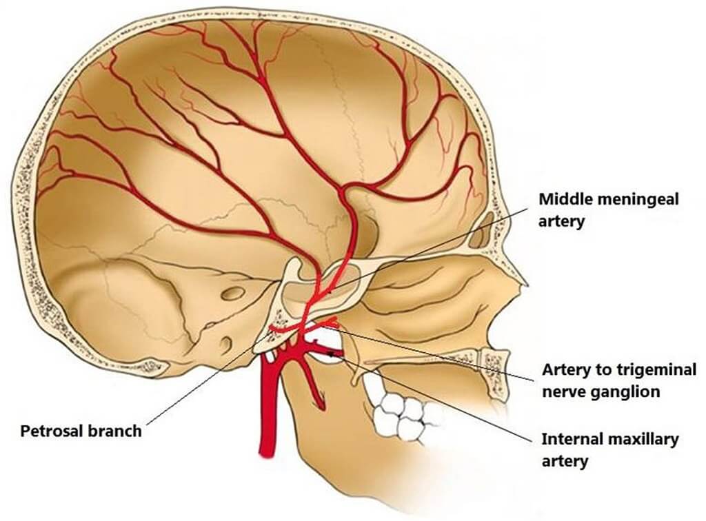
Innervation of the meninges | Radiology Reference... | Radiopaedia.org The sensory innervation of the meninges is primarily by meningeal branches of both the trigeminal and vagus nerves with a smaller contribution from the upper cervical spinal nerves 1,2. The supratentorial dura mater is mainly supplied by the ophthalmic division of the trigeminal nerve 3. Like...
Meninges Explained - Anatomy 101 For Patients
CT Brain Anatomy - Meninges Tutorial on the anatomical location of the meninges relating to acute CT brain pathology. The falx cerebri and the tentorium cerebelli are thick infoldings of the meninges which are visible on CT imaging. Elsewhere the meningeal layers are not visible on CT as they are closely applied to the...
Meninges - Nervous System

Cranial Meninges Structure infographic - LifeMap Discovery
:max_bytes(150000):strip_icc()/meninges-56f99a4f5f9b5829866fe6a7.png)
Meninges: Function and Layers, and Health Problems

the meninges of the brain Diagram | Quizlet

Anatomy of the Nervous System | Microbiology

Meninges and Cerebrospinal Fluid (Gross Anatomy of the Brain ...
Meninges Brain Labeled: Stock-Vektorgrafik (Lizenzfrei) 24964390

Meninges of the brain art print poster
Meninges, Ventricles, CSF and brain blood supply | Kenhub

Meningeal layers, the SAS, the pia mater, and the arachnoid ...
Anatomy of the Brain and Meninges Stock Photo - Alamy

408 Meninges Stock Photos, Pictures & Royalty-Free Images ...

Meninges of the brain - labeled. | CanStock

Dura mater - Wikipedia

Meninges Layers, Function & Anatomy | Dura Mater, Arachnoid ...

Brain meninges, illustration - Stock Image - C029/9056 ...
Meninges: What They Are & Function

Gray's Anatomy Meninges Dura mater Cerebellar tentorium ...
Meninges of the Brain - Adult
Anatomy of the Brain and Meninges Stock Photo - Alamy

Figure, Brain meninges. Image courtesy O. Chaigasame ...
Dura mater - Wikipedia

1: Anatomy of the human head showing the scalp, skull ...

Meninges | Radiology Reference Article | Radiopaedia.org
Definition of meninges - NCI Dictionary of Cancer Terms ...
0 Response to "39 meninges of the brain diagram"
Post a Comment