42 Horse Leg Anatomy Diagram
Dog Leg Anatomy Explained: Injury Types And Treatments ... Understanding and knowing your dog's leg anatomy will help learn the possible weaknesses, injuries, and the best ways how to treat them. The dog is carried around by the forelegs and the hind legs. Much as the hind legs have got larger muscles which make them stronger, they only carry around one-third of its body weight. PDF How Do We Diagnose Lameness in Your Horse STIFLE ANATOMY - diagram This diagram (lateral or side view of the stifle) shows important soft tissue structures not visible on radiographs. It depicts the medial or inside of the stifle, and shows the medial collateral (femoro-tibial) ligament, the medial meniscus, and two of the patetellar ligaments (the horse has three).
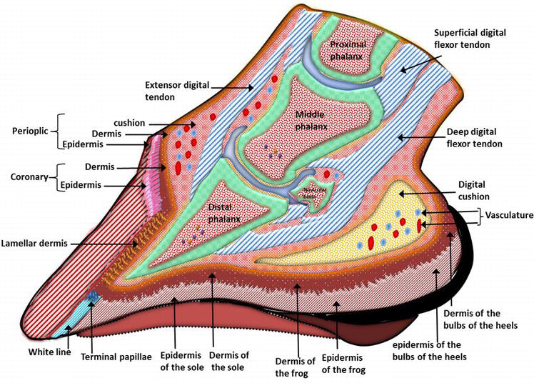
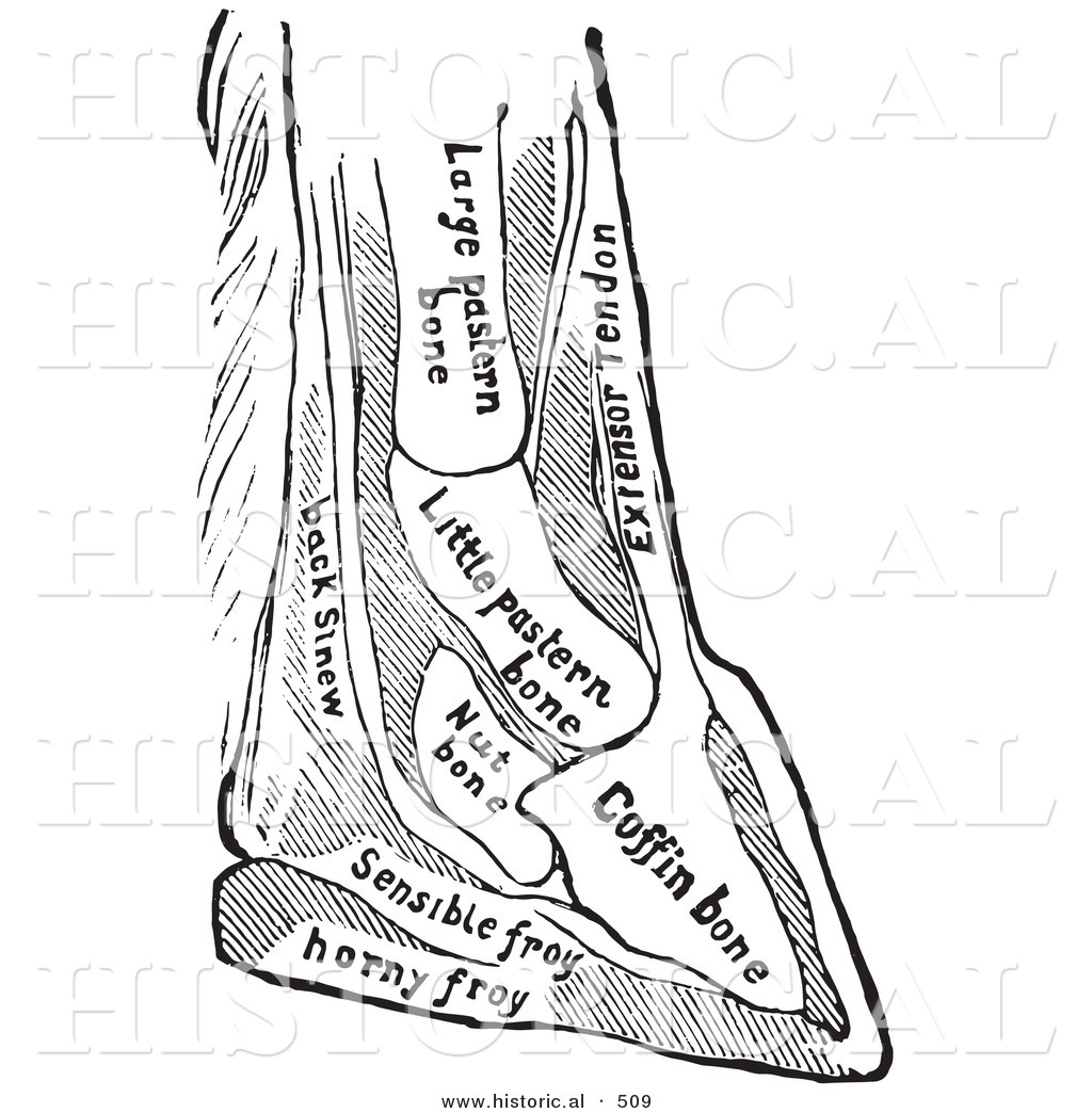
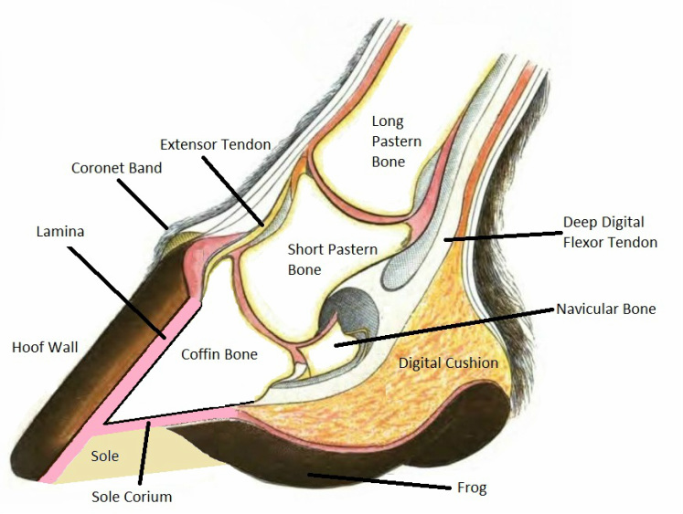
Hoof Anatomy - A Beginner's Guide - The Equine Podiatry ... The hoof is heavily supplied with blood through the two arteries which run down the back of the leg and into the foot. The pedal bone itself has an unusually high density of blood vessels within it. The photograph shows the laminae which keep the hoof wall tightly bonded to the internal structures. ... "Anatomy of the Horse", Klaus-Dieter ...
Horse leg anatomy diagram
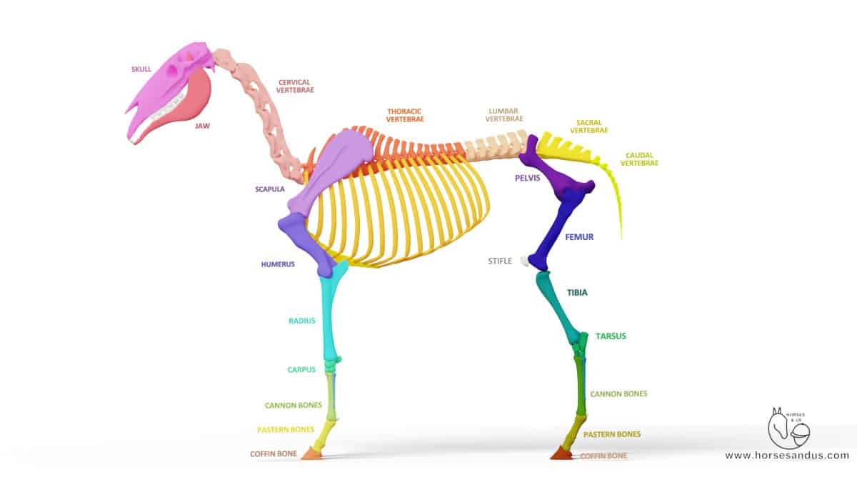
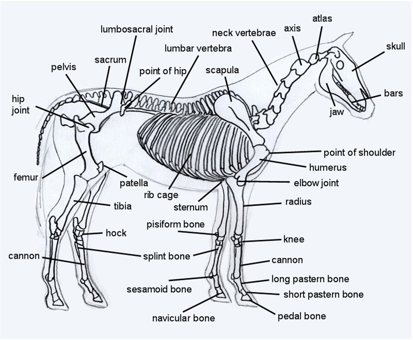
› en › libraryPerineal region: Anatomy, definition, diagram | Kenhub Feb 14, 2022 · The ischioanal fossa occupies most of the anal triangle. In a transverse section through the pelvis (in a lithotomy position), it has a horse shoe appearance, while in a coronal section (through a vertically erect individual) each fossa appears roughly pyramidal. The ischioanal fossa is limitted and comprised of the following structures: Leg Muscle Anatomy, Function, & Diagrams | Leg Muscles ... Leg Muscle Anatomy The legs are the lower limbs of the human body that provide support and stability in addition to allowing movement. The legs include the upper leg, knee, lower leg, ankle, and foot. › horse-skeletonHorse Skeleton Diagram - EquineSpot.com Take a look at this drawing of a horse skeleton. You are looking at about 205 bones that make up the equine skeletal anatomy. The more you study this picture the better understanding you will have of how a horse is built and how he moves.
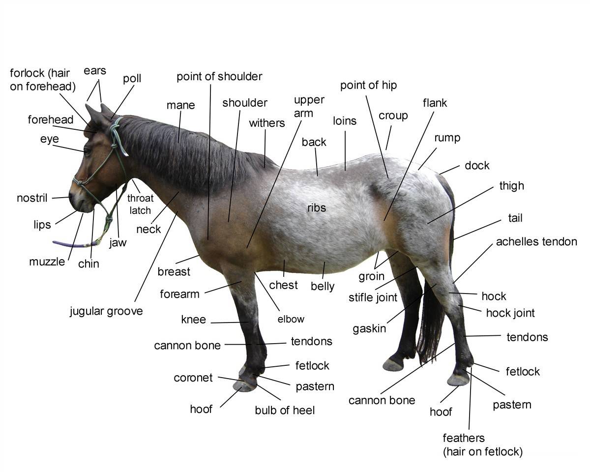
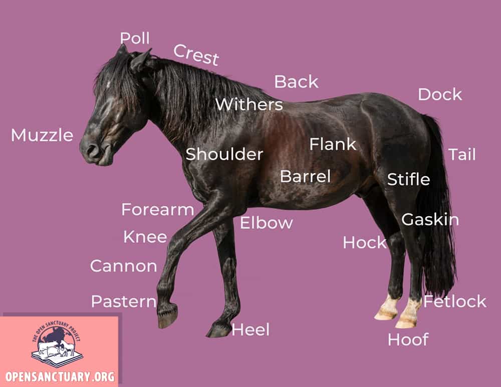
Horse leg anatomy diagram. 15 Best Images of Horse Anatomy Worksheets Printable ... Talking about Horse Anatomy Worksheets Printable, we have collected some related images to complete your references. horse body parts diagram, horse skeleton diagram and animal nervous system diagram are some main things we want to present to you based on the gallery title. Horse Leg Anatomy - Front and Rear Leg Anatomy The horse leg anatomy in the rear includes the bones of the pelvis (the ilium, ischium and pubic bones), femur, tibia, fibula, metatarsus and the phalanxes. It also includes the joints of the hip, stifle, hock, fetlock, pastern, and coffin #19-The stifle is the largest single joint in the body. Horse anatomy - diagrams of horse body parts - EQUISHOP ... Occiput - is right behind the ears, exactly where the bridle comes (namely, the occiput strap of the bridle), it is a part that joins the horse's head with his neck. Crown - is between the ears and in front of them. It is a part, which lines are marked by the eye fovea, temples with the temporal fovea, forehead, eye sockets, and eyes. Basic Horse Anatomy: Part 1 - The Open Sanctuary Project The hoof is the foot of the horse, consisting of a hard exterior and softer interior. Similar to a fingernail but much stronger. Knee. The knee (carpus) is a large, bending joint of the front leg. It functions more like a wrist than a knee. Muzzle. The muzzle consists of the nose, mouth, and chin of a horse. Pastern.
› digestive-disorders › digestive-systemThe Digestive System Diagram, Organs, Function, and More The esophagus is a muscular tube extending from the pharynx and behind the trachea to the stomach.Food is pushed through the esophagus and into the stomach by means of a series of contractions ... This is a detailed diagram of a horse's hoof. This will ... Mar 2, 2017 - This is a detailed diagram of a horse's hoof. This will help me apply this to my integument presentation. Mar 2, 2017 - This is a detailed diagram of a horse's hoof. This will help me apply this to my integument presentation. ... Leg Anatomy. Horse Anatomy. Animal Anatomy. Muscle Anatomy. Anatomy Bones. Horse Care Tips. Horse ... anatomylearner.com › dog-leg-anatomyDog Leg Anatomy with Labeled Diagram - Bones, Joints, Muscles ... Dog leg anatomy. First, you might have a basic idea of the different bones of the forelimb and hindlimb of a dog. Now I will provide you the few information on the other bones of dog leg anatomy with their unique features. The front leg of a dog consists of the clavicle, scapula (arm), radius and ulna (forearm), carpals, metacarpals, and phalanges (forepaw). en.wikipedia.org › wiki › Spider_anatomySpider anatomy - Wikipedia The anatomy of spiders includes many characteristics shared with other arachnids.These characteristics include bodies divided into two tagmata (sections or segments), eight jointed legs, no wings or antennae, the presence of chelicerae and pedipalps, simple eyes, and an exoskeleton, which is periodically shed.
Horse Diagram - The Main Body Parts of a Horse - Seriously ... A balanced and proportionate body is key to proper leg structure and the overall quality of a horse. As you can see in the horse diagram above a proportionate horse is usually square. A square frame means that the length of the body is equal to the height from the withers to the ground. PDF Horse Hoof and Leg Anatomy: A Guided Tour Horse Hoof And Leg Anatomy: A Guided Tour Scott J. Duggan Livestock Extension Faculty. Today's Mission Be able to visualize the skeletal anatomy of the lower leg and hoof of the horse. Develop an understanding of the causes of equine lameness and methods of treatment. Parts of the Horse. No Hoof, No Horse. 18 Best Images of Leg Anatomy Worksheets - Lower Limb ... Beside that, we also come with more related ideas as follows free printable human anatomy coloring pages, lower leg muscle diagram blank and lower limb bones unlabeled. Our goal is that these Leg Anatomy Worksheets pictures gallery can be a direction for you, bring you more references and also make you have a great day. Basic Horse Anatomy for Equine Owners - EquineSpot.com Basic Horse Anatomy for Equine Owners. Get the basics on horse anatomy that every horse owner needs. Diagrams, illustrations and charts will help you understand how your horse is put together. From equine skeletal anatomy to body parts and teeth. Develop a better understanding of where leg injuries occur, and the inner workings of the horse hoof.
Horse Anatomy - Complete Guide to Learn Anatomical Features ...
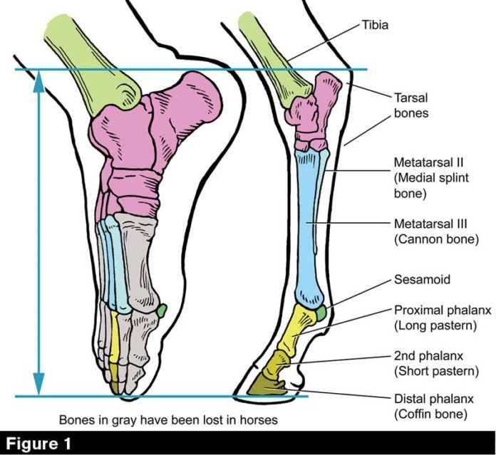
Horse Leg Anatomy - Learn Everything You Did Not Know ... The horse leg anatomy in the rear includes the bones of the pelvis (the ilium, ischium and pubic bones), femur, tibia, fibula, metatarsus and the phalanxes. It also includes the joints of the hip, stifle, hock, fetlock, pastern, and coffin. Hind limbs
Horse Anatomy - Complete Guide to Learn Anatomical Features ...
Horse Anatomy - Mobility Health Each hind limb of the horse runs from the pelvis to the navicular bone. The bones and the joints in between include: Femur (thigh) Patella Stifle joint Tibia Fibula Tarsal (hock) bone and joint Large metacarpal Small metacarpal Fetlock joint Pastern joint Coffin joint
Hock Provides the Horse Thrust Under Immense Strain
Groin Muscle Injuries - Anatomy | Dr. Mel Newton View from the back of the horse. Note that the sartorius isn't visible since it's more forward on the inside of the leg. Now we have cutaway the horse to the point where we can see the inside of the thigh. In this diagram the tail is towards the left and the head is on the right. Note that the red gracilis m. covers most of the inner surface.

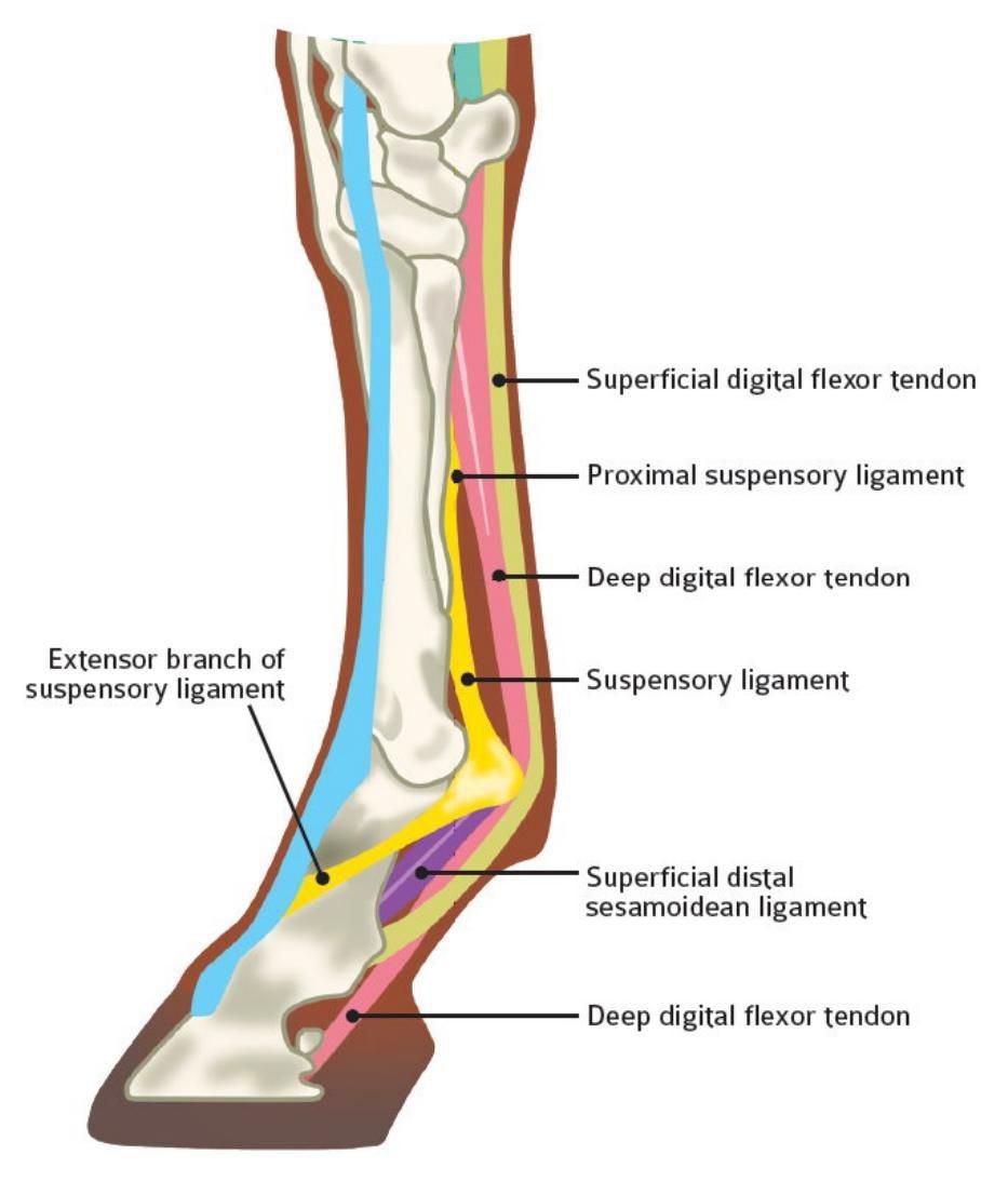
Anatomy of the distal forelimb of the horse. (a) The fetlock ...
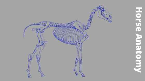
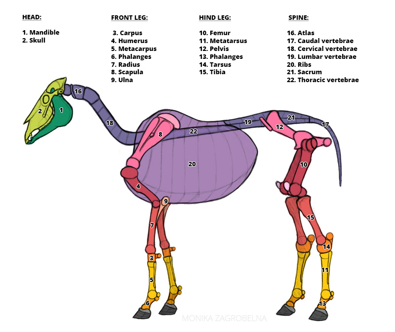
Horse Anatomy - Skeleton & Anatomy Diagram Of A Horse Horses have, on average, a skeleton of 205 bones. A significant difference in the bones contained in the horse skeleton, as compared to that of a human, is the lack of a collarbone. Their front limb system is attached to the spinal column by a powerful set of muscles, tendons and ligaments that attach the […]
Equine Anatomy — Burlington Equine Veterinary Services
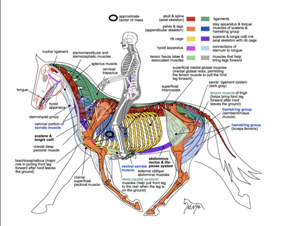
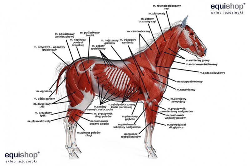
Horse Anatomy Diagrams - Directional Terms, Skeleton, and ... There are many possible diagrams of the anatomy of horse tissues. This is because there are many layers of muscles. This diagram shows the superficial layer of the tissue.. Superficial - More external or towards the surface of the body.. In this picture it shows the muscles that are closest to the surface of the skin, making them superficial.. Deep- More internal, or towards the center of the ...

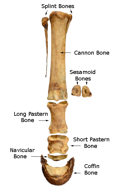
Equus distal forelimb skeletal anatomy. (a) Middle and distal ...
Horse Anatomy Pictures-Think Like a Horse-Rick Gore ... Horse Anatomy Pictures: The Amazing Horse: This page is mostly photos, graphs and charts about the horse. Some photos appear to be close to the same, but all explain some things differently. I hope it improves your understanding of the physical horse. Click the 100% button at the bottom right of this screen to enlarge pictures.
Functional Anatomy of the Horse Foot | MU Extension
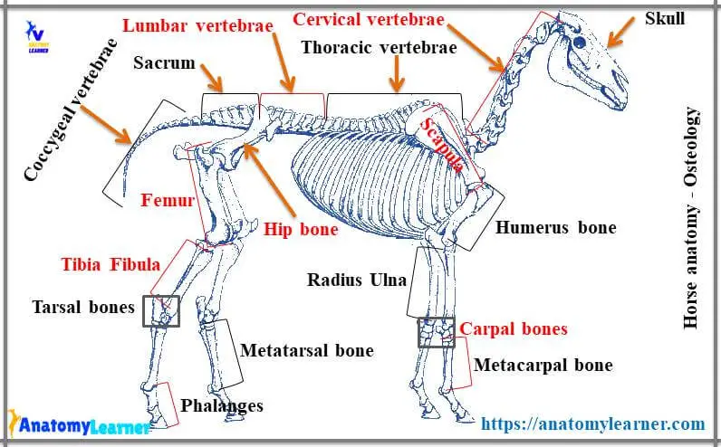
Anatomy of the horse: osteology - vet-Anatomy - IMAIOS Equine anatomy - Illustrated atlas of the bones of the horse. This module of vet-Anatomy presents 135 labeled anatomical illustrations of the osteology of the horse, specially illustrated and selected for veterinary students and equine veterinarians.
The Anatomy, Histology and Physiology of the Healthy and Lame ...
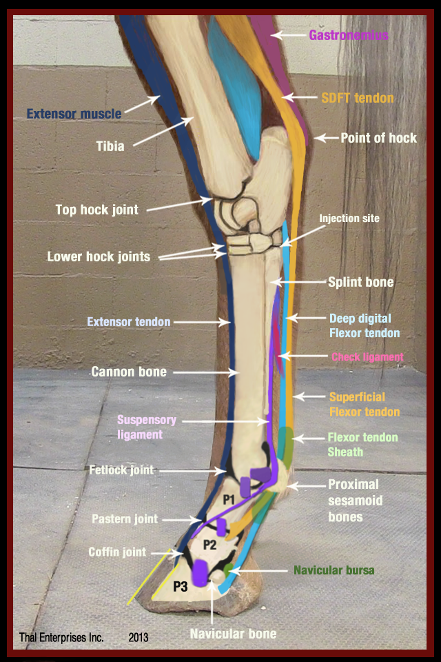
Equine Tendons & Ligaments Quiz - PurposeGames.com The tendons and ligaments on the lower leg of the horse. I deal to help those studying a Level 3 diploma in horse care or equivalent. Remaining 0. Correct 0. Wrong 0. Press play! 0%. 0:00.0. Quit. Again. This game is part of a tournament. You need to be a group member to play the tournament.

219 Horse Anatomy Illustrations & Clip Art - iStock
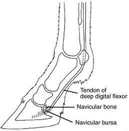
Hoof Anatomy: What Horse Hooves are Made of Hoof Anatomy: What Horse Hooves are Made of ... the deep digital flexor tendon runs down the back of the leg and wraps around the navicular bone, bending and flexing the leg. Always keep your horse's hooves healthy with regular trims, a good diet and plenty of exercise. Check your horse's hooves on a daily basis for any irregularities or ...

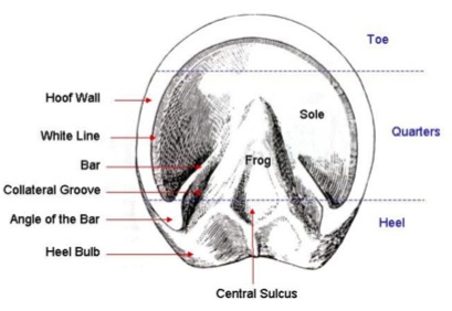
Look Into Horse Hoof Anatomy - See the Layers of the Hoof
anatomylearner.com › horse-anatomyHorse Anatomy - Complete Guide to Learn Anatomical Features ... Jul 05, 2021 · #3. Horse leg anatomy (bone and muscle anatomy) Conclusion This is the summary of horse anatomy where I tried to provide valuable information. If you think you got a basic idea of the different organs system of a horse, then share this article with your friends who are also interested to learn horse anatomy.

Horse Hind Leg Bones Horse Equus Anatomy Stock Photo ...
Horse Anatomy and Muscle Pictures - Stretch Your Horse Horse Anatomy and Muscle Diagrams. This page contains color coded pictures of the horse's hind end, deep and superficial muscles. The pictures will help you "see" which muscles you are stretching! All images are contained in the Stretch Your Horse App for free!
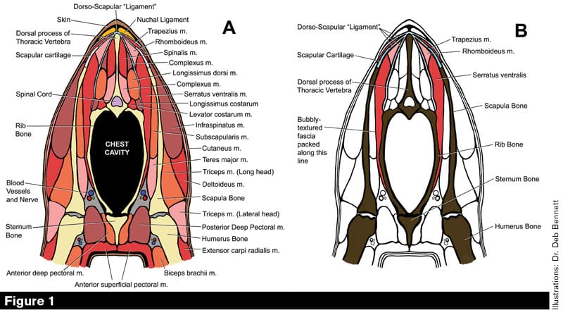
Equine Reciprocating Systems: Examining the Shoulder to ...
› horse-body-partsDiagram of Horse Body Parts - EquineSpot.com With each step the horse takes, the blood that has been forced down the leg is now forced back upward toward the heart. The front legs of the horse bear most of the horse’s weight, while the powerful back legs act as the motor that drives the animal forward. Horses carry the weight of a rider on their backs just behind the withers.

The Anatomy of Dressage Horse Hindquarters - Expert advice on ...
The 4 Basic Horse Gaits Explained [Diagrams & Animations] The 4 Basic Horse Gaits Explained [Diagrams & Animations] A gait is a distinct form of movement where the limbs move in a specific rhythmic pattern at a particular speed. As the horse changes speed, he transitions to another gait. Horses can perform a large number of gaits. Some gaits are unique to certain breeds, some gaits are acquired ...
Hoof Anatomy - A Beginner's Guide - The Equine Podiatry ...
Horse Leg Anatomy - Form and Function | EquiMed - Horse ... A horse with good conformation is going to have well-formed, symmetrical legs. When the horse is viewed from the front, the observer can drop an imaginary line from the top center of the leg at chest level down through the forearm, knee, cannon, and fetlock to the center bottom of the hoof.

Bones of the foreleg and hind leg of horse Diagram | Quizlet
Horse Leg Anatomy Photos and Premium High Res Pictures ... Browse 186 horse leg anatomy stock photos and images available, or start a new search to explore more stock photos and images. horse's foot, wood engraving, published in 1883 - horse leg anatomy stock illustrations. skeleton of the horse - horse leg anatomy stock illustrations. diagram of a horse's leg and foot - horse leg anatomy stock ...
Basic Horse Anatomy for Equine Owners
› horse-skeletonHorse Skeleton Diagram - EquineSpot.com Take a look at this drawing of a horse skeleton. You are looking at about 205 bones that make up the equine skeletal anatomy. The more you study this picture the better understanding you will have of how a horse is built and how he moves.
Parts Of A Horse - A Complete guide With 3D Visible Horse
Leg Muscle Anatomy, Function, & Diagrams | Leg Muscles ... Leg Muscle Anatomy The legs are the lower limbs of the human body that provide support and stability in addition to allowing movement. The legs include the upper leg, knee, lower leg, ankle, and foot.

Wedges, Rounded Toes, and Backward Shoes Ease Hoof Breakover ...
› en › libraryPerineal region: Anatomy, definition, diagram | Kenhub Feb 14, 2022 · The ischioanal fossa occupies most of the anal triangle. In a transverse section through the pelvis (in a lithotomy position), it has a horse shoe appearance, while in a coronal section (through a vertically erect individual) each fossa appears roughly pyramidal. The ischioanal fossa is limitted and comprised of the following structures:
Historical Vector Illustration of a Horse Diagram Featuring ...
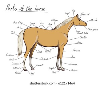
Parts Horse Equine Anatomy Equestrian Scheme Stock Vector ...
Horse anatomy - diagrams of horse body parts - EQUISHOP ...
Basic Horse Anatomy: Part 1 - The Open Sanctuary Project
Everything You Need to Know About Laminitis — Irongate Equine ...

Basic equine anatomy - The Brook Vet
Horse Anatomy for Artists: Skeleton and Muscle Diagrams ...
Horse Skeleton Diagram

Hands-on: A Guided Tour of Equine Anatomy – Focus on the Hoof ...

Horse Foot Health

Horse Leg Bones Anatomy Collection Horse Leg Anatomy Pictures ...

Limbs of the horse - Wikipedia

Equine Foot and Hoof Anatomy Chart Horse
The Distal Limb Bones of the Equine

Forever Horses: Anatomy of the Equine Hindleg

Equine Distal Limb Anatomical illustration

Horse leg muscles and skeleton structure diagram

horse leg bones Diagram | Quizlet
Horse leg detailed | | agupdate.com
Vitals & Anatomy - Horse Side Vet Guide

Equine Muscular Anatomy Chart / Poster - Laminated

Equine Internal Anatomy Chart / Poster - Laminated

Equine anatomy - Wikipedia

Equine Anatomy Chart
0 Response to "42 Horse Leg Anatomy Diagram"
Post a Comment