38 eye cross section diagram
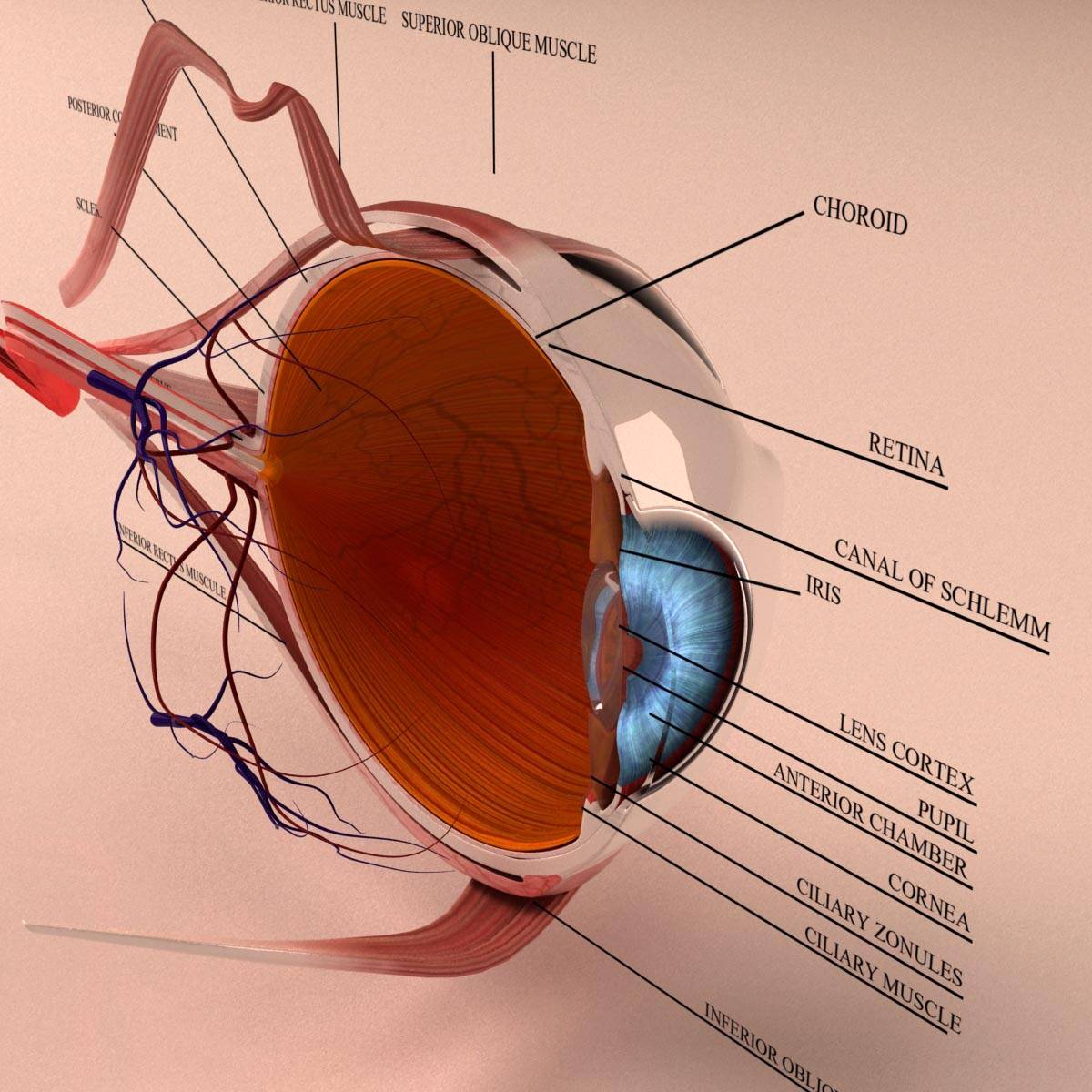
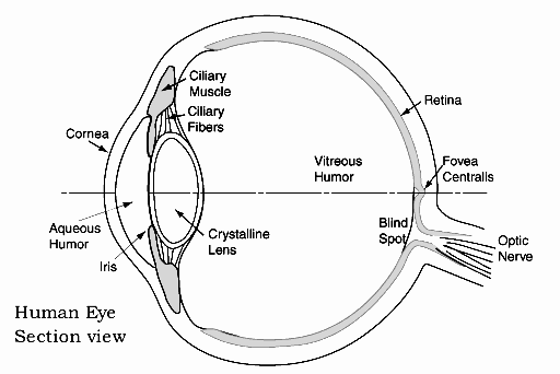
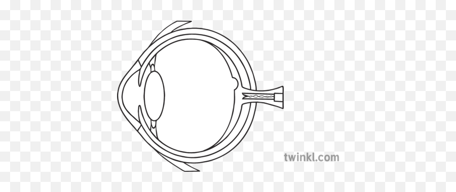
Q1.€€€€€€€€€ (a)€€€€ The diagram shows the cross-section of an eye. Use words from the box to label the parts, A, B and C. € (3) cornea€€€€€€€€€ iris€€€€€€€€€€ lens€€€€€€€€€ pupil€€€€€€€€€€€€ retina Eye Model Labeled Bing Images Eye Anatomy Diagram Eye Anatomy Diagram Of The Eye. Human Eye Anatomy 3d Model Cross Section With All Eye Parts Ready For Renderings 3d Printings Illustrations Human Eye Eye Anatomy Anatomy Models. Image Result For 3d Model Of Human Eye School Project Eye Anatomy Anatomy Models Science Models.
This resource from ABPI Schools shows the cross section of an eye. Creative Commons "NoDerivatives" Review. 4 Something went wrong, please try again later. ... report. 4. Accurate labelled diagram of the eye, showing the relevant structures with their functions. Possibly too much information on the diagram? Empty reply does not make any sense ...

Eye cross section diagram
Find Eye Cross Section Labeled Diagram stock images in HD and millions of other royalty-free stock photos, illustrations and vectors in the Shutterstock collection. Thousands of new, high-quality pictures added every day. Cross section of the eye The cornea is the clear, transparent front layer of the eye through which light passes The iris gives our eyes colour and it functions like the aperture on a camera, enlarging in dim light and contracting in bright light. Cross section of the sense organ with all important components like lens, pupil, eye chamber, retina, optic nerve and rainbow skin. Internal Parts of the Human Eye, cross-section showing the cornea, iris, lens, and retina, vintage engraved illustration.
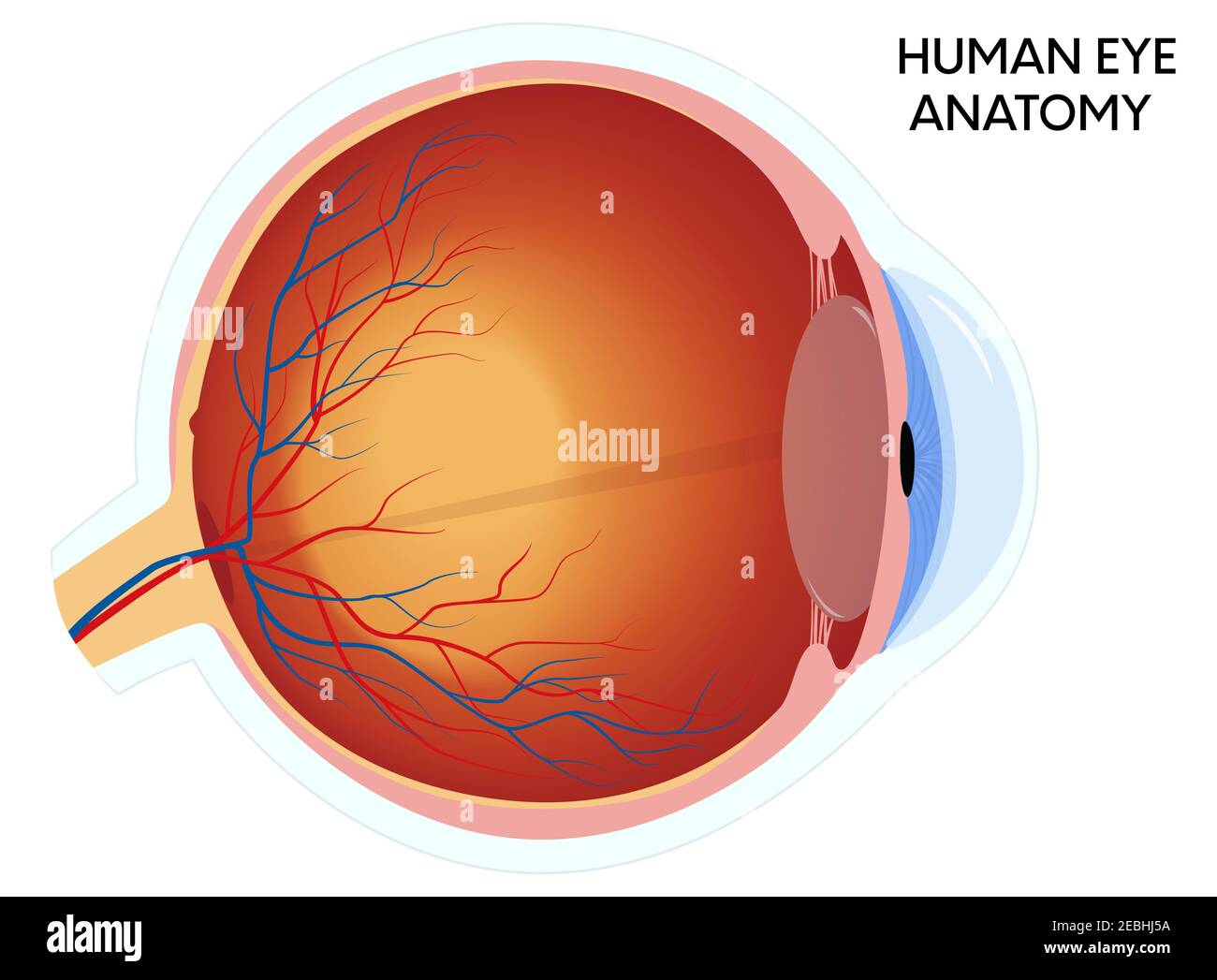
Eye cross section diagram. Cross-Section of the Human Eye Right Part anatomy educational diagram. Human eyeball section special visual educational structure for school, university, college, and hospital internship education. Human organ that reacts to light and color differentiation and generate the human vision. Müllers muscle: Sympathetically-innervated muscle extending from levator palpebrae superioris to top of tarsal plate. Elevates eyelid, but not nearly as much as levator does. Impaired in Horner Syndrome, but ptosis is mild because Müller's muscle is a weak elevator. Orbital septum: Collagenous curtain connecting frontal bone and upper lid tarsus. Superstar. Shutterstock customers love this asset! Related keywords. eye human anatomy cross eyeball section ball diagram cornea drawing iris medical vector anatomical body care college cut education eyebrows focus graphic health health care illustration learning lens look medicine nerve ocular optic optical organ part portrait pupil retina school see sense sensitive sight study symbol ... Human Eye in Cross Section. Look carefully at the diagram at the top of the page. Now check out the following questions (and answers)!
Diagram showing cross section of human eye. Holding a silicone anatomy of an eye in Hand. Diagram showing parts of human eye illustration. Corneal abrasion in the human eye. eye vein system x ray angiography vector design ... Cross section of vitreous anatomy. A, Cross section diagram of the eye with emphasis on the anatomical features of the vitreous. The vitreous is most firmly attached to the retina at the vitreous base, and it also has adhesions at the optic nerve, along vessels, at the fovea, and to the posterior lens capsule. Eye Cross Section Diagram. Here are a number of highest rated Eye Cross Section Diagram pictures on internet. We identified it from obedient source. Its submitted by doling out in the best field. We admit this nice of Eye Cross Section Diagram graphic could possibly be the most trending topic later we allocation it in google benefit or facebook. Eye diagram at the crossing points of the eye and is usually measured in picoseconds for a high speed digital signal ie 200 ps is used for a 5 Gbps signal. Cross section through the maxillary sinus. The human eye is an organ that reacts to light. Contrary to popular belief the eyes are not perfectly spherical.
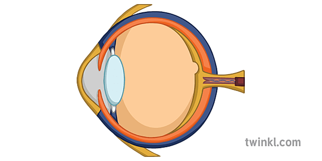
Retina Cross Section - 9 images - human eye cross section eyeball 3d model cgtrader, vegf secreted by hypoxic m ller cells induces mmp 2, Human eye cross section anatomy diagram - download this royalty free Vector in seconds. No membership needed. Human eye cross section anatomy diagram with all parts anatomical structure for medical science education and health care. | CanStock Use this human eye diagram to help you to teach children about the eye. This cross-section has been illustrated using clear black and white lines - it's ideal to use in colouring and labelling activities. The human eye has many parts that work together to produce vision: such as the pupil, iris and sclera. Cross-section anatomical diagram of a cataract developing in an eye. An eye diagram or eye pattern is simply a graphical display of a serial data signal with respect to time that shows a pattern that resembles an eye. A Cross section diagram of the eye with emphasis on the anatomical features of the vitreous. Controls forceful eyelid closure.
inner layer of posterior wall of eye (see Retina in Cross Section) contains receptors that convert light energy into signals that brain can interpret. Choroid: vascular layer that nourishes outer retina. can be inflamed in autoimmune ("rheumatologic") disorders. Sclera: collagenous outer layer of wall of eye.
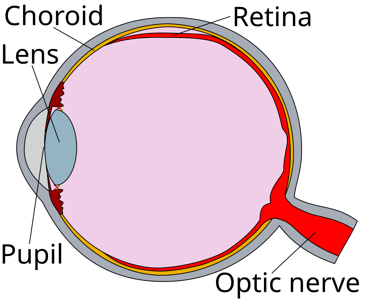
Download scientific diagram | Cross sectional diagram of human eye [1]. from publication: Identification of Diabetic Retinal Exudates in Digital Color Images Using Support Vector Machine ...
cross-section diagram of human eye. - retina diagram stock illustrations antique medical scientific illustration high-resolution: eye - retina diagram stock illustrations Diagram Of A Scleral Buckle, Used To Repair A Retinal Detachment.
Cross section of ground with grass and steel pipe with water isolated on white. House in cross-section. Modern house, villa, cottage, townhouse with shadows. Architectural visualization of a three. Isometric Cross section through a window pane PVC profile laminated wood grain, classic white. Set of Cross-section. House in cut. Detailed interior.
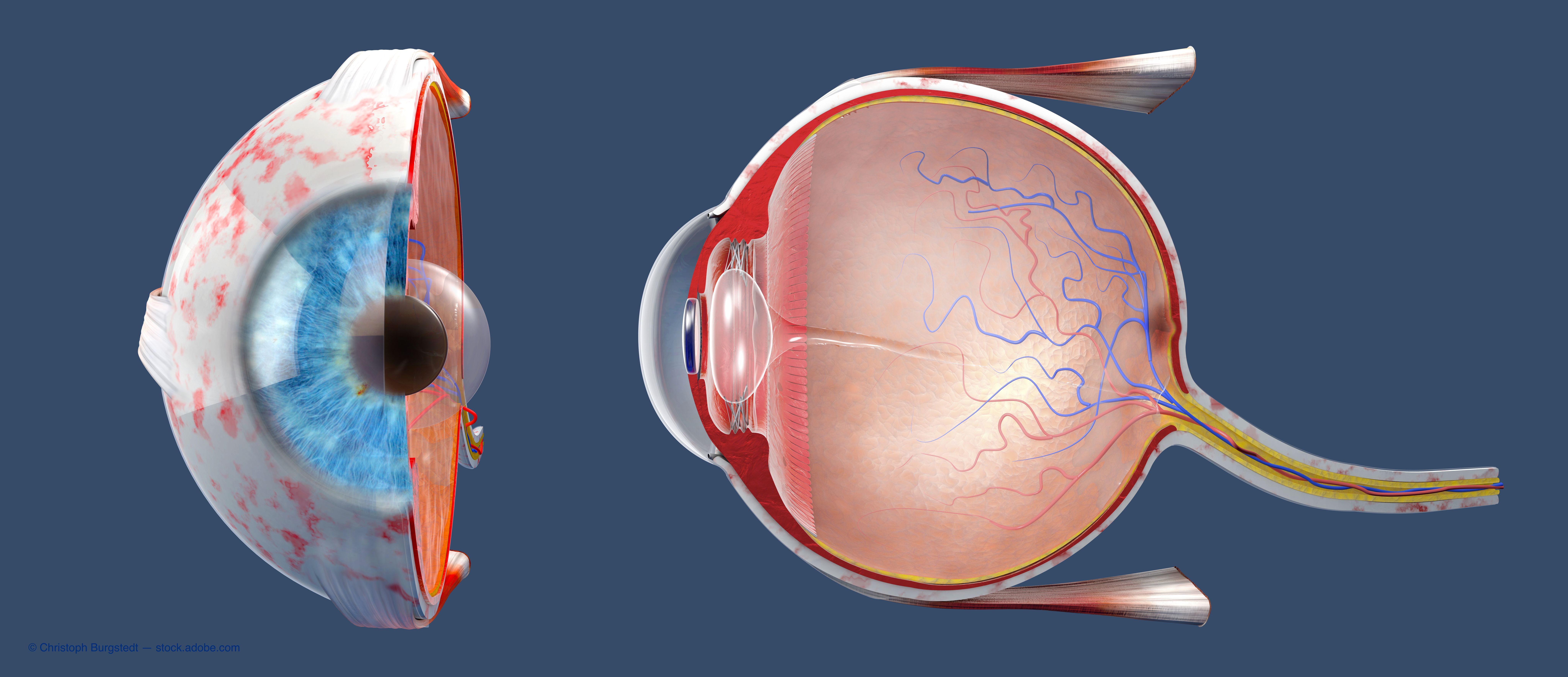
Eye Cross section Low-poly 3D model. Add to wish list Remove from wish list. Report this item. Description; Comments (0) Reviews (0) Visualization of the Cross of the eye . The illustration shows the 3 different distinct layers of the eye which are the sclera , the choroid and the retina. The lens and its suspensory ligaments are also seen .
Eye Eye cross section Eye Cross-section When light strikes the eye, the first part it reaches is the cornea, a dome positioned over the center of the eye. The cornea is clear and refracts, or...
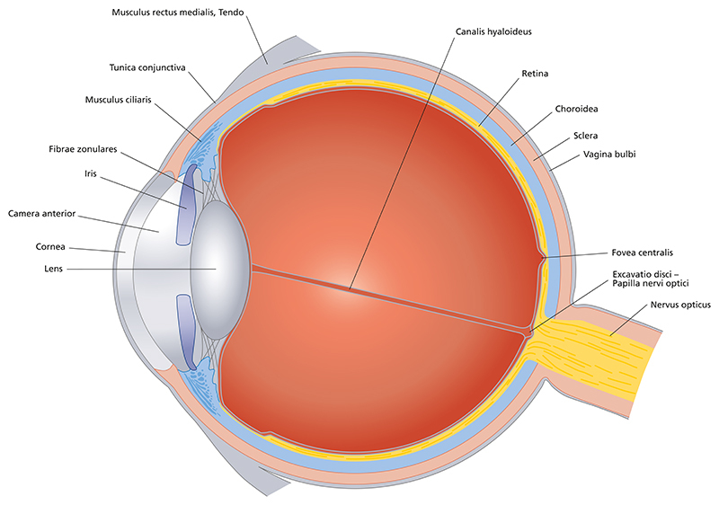
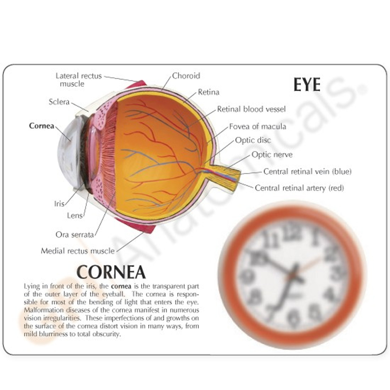
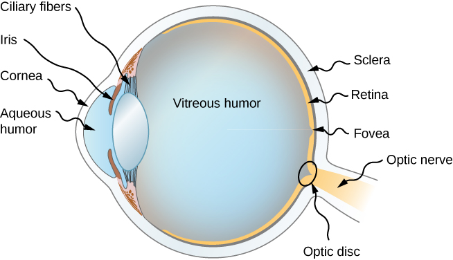
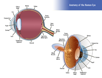
The diagrams below show cross sections of the human eyeball. As we journey through the different structures, refer to the diagrams to quickly digest the content on this page. Our eyeballs are fairly round organs cushioned by fatty tissues and they sit in two bony sockets inside the skull. This helps to protect our eyes from injury.
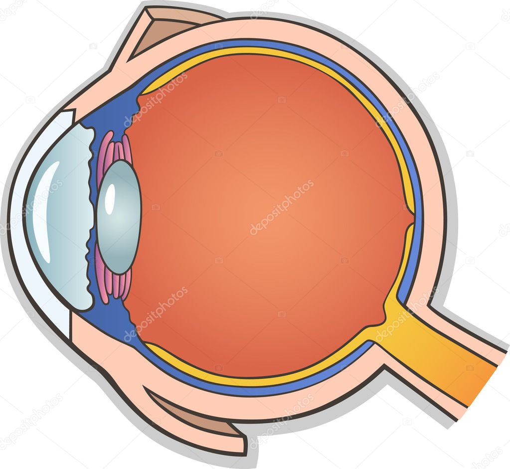
diagram of eye 1. Drawings of the Eye Cross section drawing of the eye - (side view) with major parts labeled. Cross section drawing of the eye - (rear view). 2. Cut-away view of the eye in its socket showing the: bony socket, orbital muscles, eyelids and eyelashes.
Eye anatomy blank worksheet, printable test illustrations. Brain and eye anatomical cross section diagrams. School lesson activity educational workbook page.
the outer eye as we see it with all parts labeled lateral view of the eyeball in the skull top view of the eyeball in the skull diagram of the visual field large central illustration of a lateral cross section of the eye medial cross section of the eye close up views of the: anterior chamber angle lens retina fundus macula lutea Made in the USA.
Here are a number of highest rated Eye Cross Section Diagram pictures on internet. We identified it from trustworthy source. Its submitted by dealing out in the best field. We put up with this kind of Eye Cross Section Diagram graphic could possibly be the most trending topic in imitation of we portion it in google improvement or facebook.
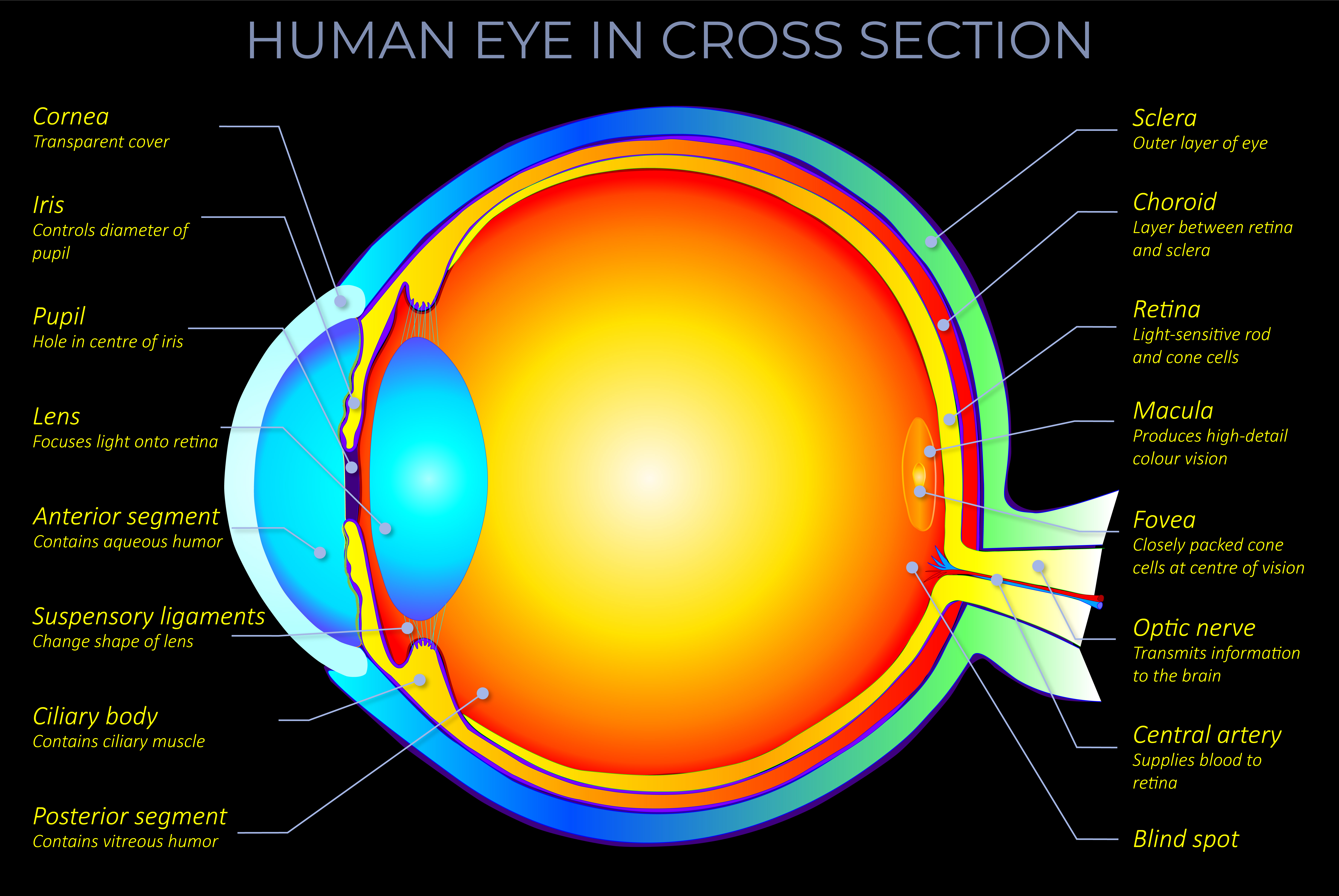
Cross section of the sense organ with all important components like lens, pupil, eye chamber, retina, optic nerve and rainbow skin. Internal Parts of the Human Eye, cross-section showing the cornea, iris, lens, and retina, vintage engraved illustration.
Cross section of the eye The cornea is the clear, transparent front layer of the eye through which light passes The iris gives our eyes colour and it functions like the aperture on a camera, enlarging in dim light and contracting in bright light.
Find Eye Cross Section Labeled Diagram stock images in HD and millions of other royalty-free stock photos, illustrations and vectors in the Shutterstock collection. Thousands of new, high-quality pictures added every day.

![Cross sectional diagram of human eye [1]. | Download ...](https://www.researchgate.net/publication/276541864/figure/fig1/AS:612895498964992@1523137082339/Cross-sectional-diagram-of-human-eye-1.png)













0 Response to "38 eye cross section diagram"
Post a Comment