40 acl and mcl diagram
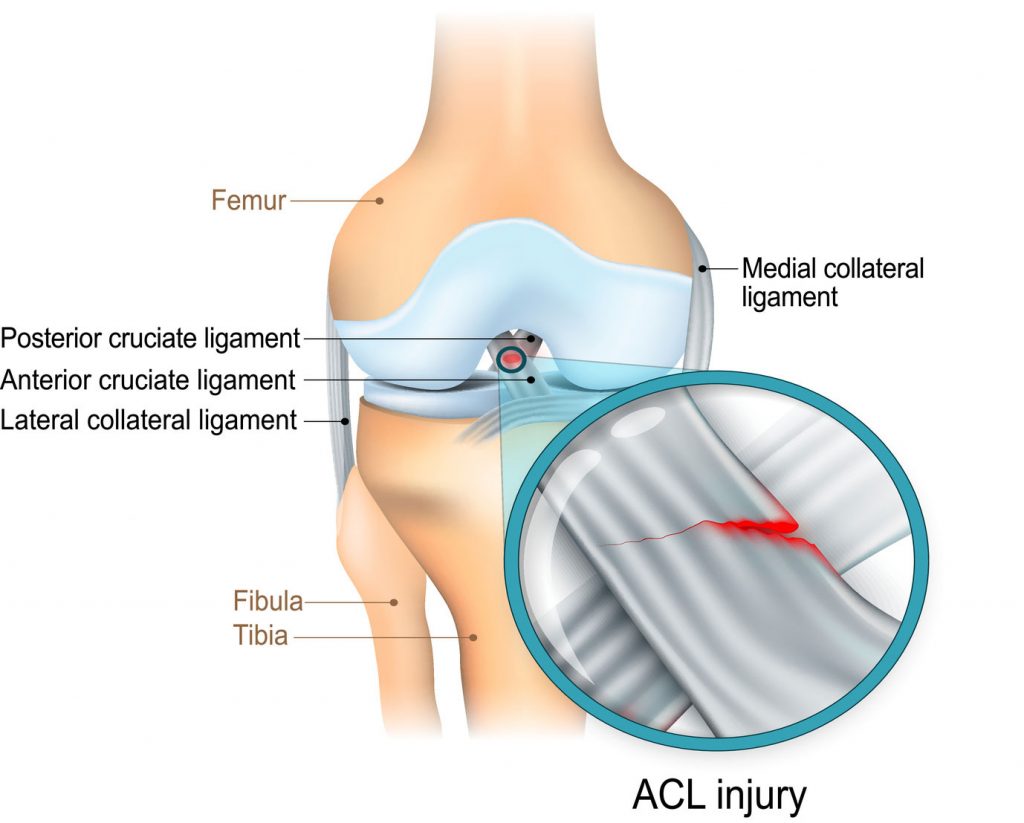
ACL (anterior cruciate ligament) strain or tear: The ACL is responsible for a large part of the knee’s stability. An ACL tear often leads to the knee “giving out,” and may require surgical ... 21.06.2021 · Anterior cruciate ligament (ACL) avulsion fracture or tibial eminence avulsion fracture is a type of avulsion fracture of the knee. This typically involves separation of the tibial attachment of the ACL to variable degrees. Separation at the femo...
23.04.2015 · Medial collateral ligament (MCL): This broad, flat, ligament is on the outside of the knee and connects the head of the femur to the head of the tibia. It is commonly injured in sports involving ...
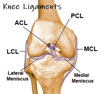
Acl and mcl diagram
... Anterior Cruciate Ligament (ACL). ACL, PCL, MCL, and LCL injuries are caused by overstretching or tearing of a ligament by twisting or wrenching the knee. 31.07.2021 · Segond fracture is an avulsion fracture of the knee that involves the lateral aspect of the tibial plateau and is very frequently (~75% of cases) associated with disruption of the anterior cruciate ligament (ACL).On the frontal knee radiograph, it may be referred to as the lateral capsular sign. Anterior to the intercondyloid eminence of the tibia, being blended with the anterior horn of the medial meniscus. The tibial attachment is in a fossa in front of and lateral to anterior spine, a rather wide area from 11 mm in width to 17 mm in AP direction.. For more detail on the anatomy of the ACL, please see this page: Anterior Cruciate Ligament (ACL) - Structure and Biomechanical Properties
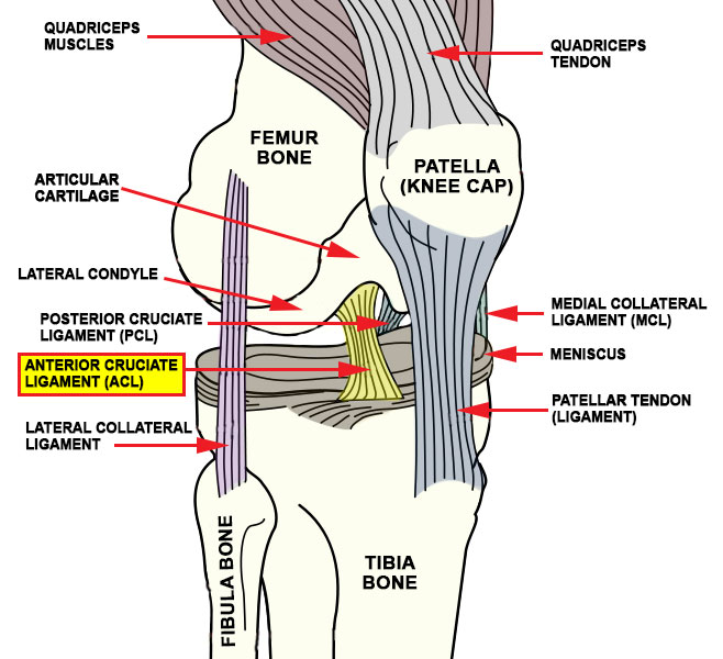
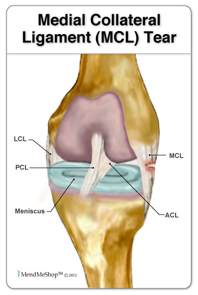
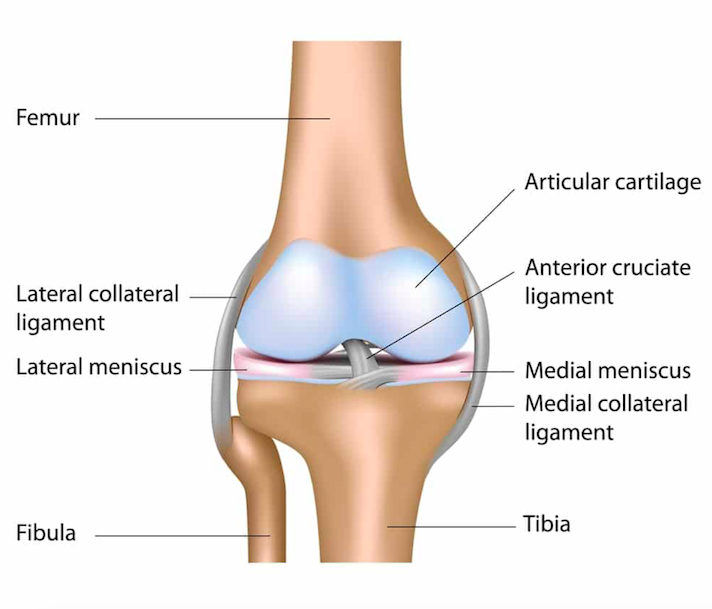
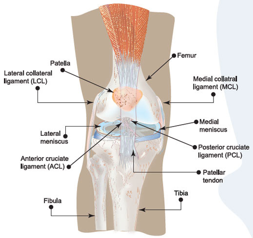
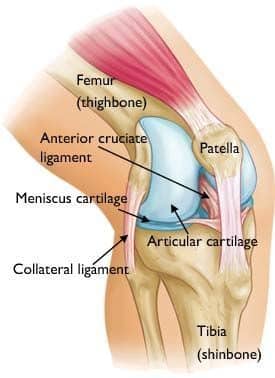
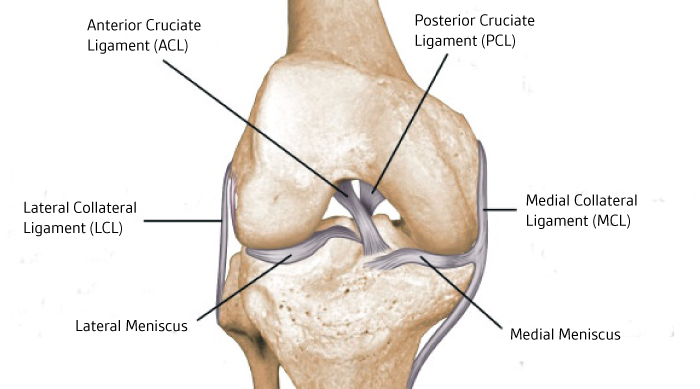
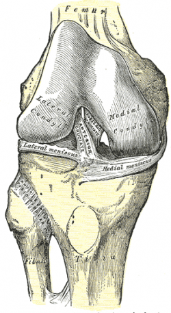
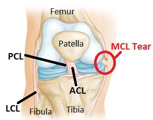
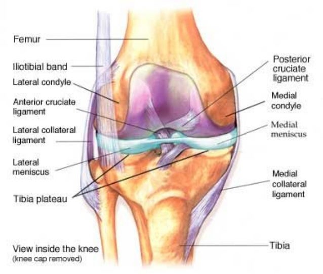
Acl and mcl diagram. An anterior cruciate ligament (ACL): Inside of the knee and crosses to the front. A posterior cruciate ligament (PCL): Inside of the knee and crosses to the back. The MCL and the LCL sit on the sides of the knee, and they help give stability to the knee if your knee gets hit from the sides. Knee bones, ligaments, and meniscus The anterior cruciate ligament (ACL) is one of a pair of cruciate ligaments (the other being the posterior cruciate ligament) in the human knee.The two ligaments are also called "cruciform" ligaments, as they are arranged in a crossed formation. In the quadruped stifle joint (analogous to the knee), based on its anatomical position, it is also referred to as the cranial cruciate ligament. These are the four main ligaments or tough bands of tissue in the knee. ACL is the anterior cruciate ligament, which connects the thigh bone to the shin bone. MCL Tear Symptoms. Just as with an ACL tear, you may hear a popping sound when the injury occurs. Some of the most common symptoms of an MCL tear include: Pain on the inner side of the knee. Feeling the knee “give out”. Swelling. The knee feels unstable. Pain when putting weight on the knee. Locked knee.
ACL vs. MCL tears: Although symptoms of ACL and MCL tears are similar, a few key differences will help identify whether the injury affected the ACL or MCL. An ACL tear will have a more distinctive and loud popping sound than an MCL tear. The location of your pain and swelling could indicate either an ACL or MCL tear. Acl And Mcl Diagram. Signs and symptoms of a medial collateral ligament (MCL) injury include The anterior cruciate ligament (ACL) and posterior cruciate ligament. The MCL is one of four major ligaments that supports the knee. either partial or complete ruptures in the ligament significantly increases the load on the ACL. 23.09.2021 · MCL tears can hurt for a while… and the pain can be severe. The diagnosis depends on the type of “injury”. If the pain came on suddenly without a fall or sports injury and you are over 55 then this could be a meniscus tear, or something we call and insufficiency fracture (stress fracture). Best to see a knee doctor to help you figure this ... 22 Mar 2018 — Knee injuries are very common among athletes, with the most dreaded of those injuries being an ACL or MCL tear. The knee is a complicated ...
The Anterior Cruciate Ligament (ACL) and Medial Collateral Ligament (MCL) are two important structures in the knee. Along with other ligaments, the ACL and MCL work together to help keep the knee stable and moving properly. A torn ACL can occur in conjunction with a Grade 1, 2, or 3 MCL tear. 25.03.2021 · Investigations. Proctoscopy can be used to visualise the opening of the tract in the anal canal. For complex fistula, MRI imaging is often required to visualise the anatomy of the tract. Park’s classification system divides anal fistulae into four distinct types (Fig. 2):. Inter-sphincteric fistula (most common) Trans-sphincteric fistula; Supra-sphincteric fistula (least common) Ligaments are strong, elastic-like tissues that connect bone to bone and provide stability and protection to your knee joint by limiting the forward and backward movement of the shin bone. There are four ligaments in the knee joint that connect the femur and tibia; the anterior cruciate ligament (ACL), the posterior cruciate ligament (PCL), the medial collateral ligament (MCL), and the lateral ... Here’s a diagram of the knee that explains what and where the various ligaments are: Anatomy of the Knee. ACL = Anterior Cruciate Ligament (this has gone on me) MCL = Medial Collateral Ligmanet (also gone) – this is on the inside of my right knee. LCL = Lateral Collateral Ligament (also gone – outside of my right knee)
30.10.2021 · That makes him overlooked rather than overrated--he's only overrated to the above Venn diagram. Click to expand... I'm not quite sure I'm smart enough to fully understand what you're saying here. Richards had some great seasons, but he was an overhyped player. Playing in Philly was part of it too. Carter for example, is the better player, and also more responsible off the ice, but he'll never ...
25 Jan 2016 — Learn the differences between ACL and MCL tears. Knee pain often indicates one of ... diagram of all parts of the knee joint and ligaments ...
Assesses: MCL + ACL (Rotator Instability) The test is performed with the patient in a relaxed, supine position. The knee to be tested should be fully extended and the hip flexed to approximately 30 degrees. One of the examiners hands holds heel of the foot of the leg to be tested. The examiner’s other hand is on the lateral aspect of the ...
Multiligament Knee Injury (ACL/PCL +/- MCL/PLC) Rehabilitation following surgery for multiligament knee injury (MLKI) or knee dislocation is an essential element of the treatment to achieve a full recovery. This protocol is intended to provide the user with instruction, direction, rehabilitative guidelines and functional goals. It is not meant
Download scientific diagram | Deep and superficial MCL, and ACL double bundle anatomy from publication: Consideration of growth factors and bio-scaffolds ...
An anterior cruciate ligament injury occurs when the anterior cruciate ligament (ACL) is either stretched, partially torn, or completely torn. The most common injury is a complete tear. Symptoms include pain, a popping sound during injury, instability of the knee, and joint swelling. Swelling generally appears within a couple of hours. In approximately 50% of cases, other structures of the ...
18.02.2018 · I had an ACL surgery on right side but when i stand on one leg left leg stabilization seems worse than right leg also left leg knee feels too much mobile compared to right (acl side). When i begin the exercises esp. left external rotation exercises, i begin feeling my left knee less mobile (or more controlled). This feels good. I know i may have swayback on my right side but the thing is ...
Anterior to the intercondyloid eminence of the tibia, being blended with the anterior horn of the medial meniscus. The tibial attachment is in a fossa in front of and lateral to anterior spine, a rather wide area from 11 mm in width to 17 mm in AP direction.. For more detail on the anatomy of the ACL, please see this page: Anterior Cruciate Ligament (ACL) - Structure and Biomechanical Properties
31.07.2021 · Segond fracture is an avulsion fracture of the knee that involves the lateral aspect of the tibial plateau and is very frequently (~75% of cases) associated with disruption of the anterior cruciate ligament (ACL).On the frontal knee radiograph, it may be referred to as the lateral capsular sign.
... Anterior Cruciate Ligament (ACL). ACL, PCL, MCL, and LCL injuries are caused by overstretching or tearing of a ligament by twisting or wrenching the knee.






/knee-pain-instability-2549493-5c04aaf946e0fb00010b8e7a-b0ef89c536aa4cd7a9db3f927d72597b.png)




:strip_icc():format(jpeg)/kly-media-production/medias/3107265/original/099932600_1587383541-ACL_1.jpg)




0 Response to "40 acl and mcl diagram"
Post a Comment