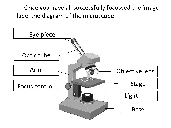
37 diagram of a microscope with labels
Compound microscope - It comes with more than one lens and provides better magnification than the simple microscope. A compound microscope is also called a bright field microscope. It can provide magnification by up to 1,000 times. Stereo microscope/dissecting microscope - It can magnify objects by up to 300 times. It is used to visualize ... Light microscope, optical microscope diagrams. Label microscope diagram. Microscope labeled diagram. Microscope lens. Elizabeth Mims2019-2020 ...
Microscope, Microscope Parts, Labeled Diagram, and Functions Published by Admin on June 1, 2021 June 1, 2021. What is Microscope? Microscope is derived from Ancient Greek words and composed of mikrós, "small" and skopeîn,"to look" or "see".

Diagram of a microscope with labels
11. Obtain a prepared microscope slide of a nerve. Locate the cross section of the nerve, and note the many round nerve fibers inside. Nerve fiber is a. Figure 25.1 Label this diagram of a motor neuron. Figure 25.1 Label this diagram of a motor neuron. Figure 25.2 Label the features of the myelinated nerve fiber. 40x 400x Compound Monocularbiological Microscope45 Degree Angled Headelectric Lightedbeginner Slides Plant Cell Things Under A Microscope Plant Cell Picture. Plasmolysis Of Elodea Biology Experiments Apologia Biology Teaching Science. Elodea Leaf Cell Under Microscope Plant Cell Cells Worksheet Lab Activities. Compound Light Microscope Diagram. A Study Of The Microscope And Its Functions With A Labeled Diagram. Free Microscope Drawing Download Free Clip Art Free Clip Art On. Light Microscope Sketch At Paintingvalley Com Explore Collection. Compound Microscope Drawing Clipart Library Clip Art Library.
Diagram of a microscope with labels. View Label By Identifying The Parts Of A Microscope Using The Word Pictures. Use page r8 in the back of your textbook to label the diagram below. Label the parts of the microscope with answers (a4) pdf print version. Labeled microscope worksheet answers. High power objective 6. Students label the parts of the microscope in this photo of a basic laboratory light microscope. Each microscope layout both blank and the version with answers are available as pdf downloads. When focusing a specimen you should always start with the objective. Animal Cell Diagram Under Electron Microscope. angelo. October 8, 2021. 25 Animal Cells Diagram With Labels Markcritz Template Design Cell Diagram Cell Diagram Project Plant And Animal Cells. Sel Tumbuhan Dan Sel Hewan Cell Organelles Animal Cell Cell Diagram. Pin By Simparinka Samuel On Bikotwa Junior Cell Diagram Plant Cell Diagram Plant Cell. Each microscope layout (both blank and the version with answers) are ... Download the Label the Parts of the Microscope PDF printable version here.
Microscope Labeled Diagram. Revision Questions : FAQ. How to tell the difference between a standard condenser and an Abbe condenser? Using a condenser, the illuminator's light can be collected and focused upon a specimen. Under the microscope stage, near the diaphragm, they can be found. They are critical in obtaining crisp, clear images at ... Download Clker's Microscope With Labels clip art and related images now. ... A diagram showing all of the parts of a compound light microscope. These labeled microscope diagrams and the functions of its various parts, attempt to simplify the microscope for you. However, as the saying goes, 'practice makes perfect', here is a blank compound microscope diagram and blank electron microscope diagram to label. Download the diagrams and practice labeling the different parts of these. Microscope Diagram and Quiz. A collection of microscope diagrams and worksheets for science class. Download them all in one convenient PDF, and select the ...
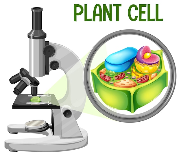
Compound Light Microscope Diagram Medical Laboratory Science. Amazon Com Swift Kids Sw50 40x 400x Magnification Beginner. Cell Structure Biology Quiz. Labelled Diagram Of Compound Microscope. A Study Of The Microscope And Its Functions With A Labeled Diagram. Biology Term 1 Diagram Quizlet. These labeled microscope diagrams and the functions of its various parts, attempt to simplify the microscope for you. Source: www.worksheeto.com Microscope , instrument that produces enlarged images of small the magnifying power of a microscope is an expression of the number of times the object being optical microscopes can be simple ... Compound microscope- definition, labeled diagram, parts, uses Compound Microscope Definition The term microscope can be split into two separate words, 'micro' and 'scope', where the term 'micro' means small or tiny, and 'scope' means to view or to observe. Plant Cell Diagram Under Electron Microscope. It's a thin slice: Here's a diagram of a plant cell: The diagram is very clear, and labeled; but at the same time it is interpretive. Though we cannot see everything through the light microscope, some important organelles are visible and we can begin to see some structural differences.
Draw A Neat Labelled Diagram Of A Compound Microscope Class 12 Physics Cbse
11+ Labeled Diagram Of Electron Microscope Pics. These labeled microscope diagrams and the functions of its various parts, attempt to simplify the microscope for you. The samples are scanned in the vacuum and so they require special preparation. In addition to optical microscopes, also have your students look at electron microscopes, which ...
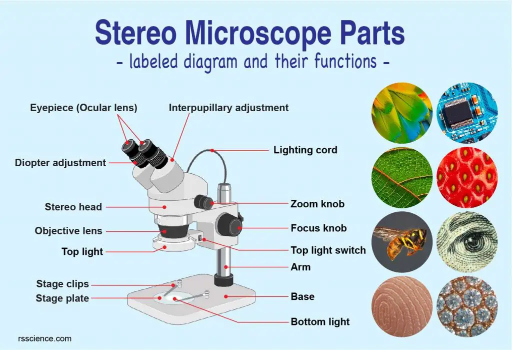
Parts Of Stereo Microscope Dissecting Microscope Labeled Diagram Functions And How To Use It
[In this figure] A collection of objective lenses. Each objective has its information (i.e. magnification) and color-code label on the side. Photo credit: Accu- ...

Microscope Banner Background Images Microscope Downloadable Resume Template
Thick and thin skin histology labeled diagram. I would like to show you the different histological features from both thick and thin skin histology slides with a labeled diagram. I hope these skin microscope slide labeled diagrams might help you to identify and learn all the structures.
1
Microscope parts and use worksheet answer key along with labeling the parts of the microscope blank diagram available for worksheet january 13 2018 we tried to locate some good of microscope parts and use worksheet answer key along with labeling the parts of the microscope blank diagram available for image to suit your needs.

Microscope Crossword With Diagram Printable Distance Learning Options
Label a compound light microscope diagram binocular microscope definition of binocular microscope by. If you are using a graticule slide a microscope slide with millimeter grid lines lightly sketch a grid over your circle. Microscopy from www2.nau.edu. Draw and label a light microscope. Observe the skin cells under both low and high power of ...
Hanovertwpschools Com
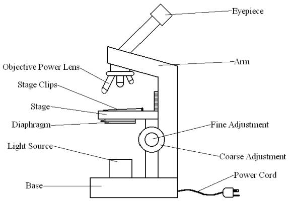
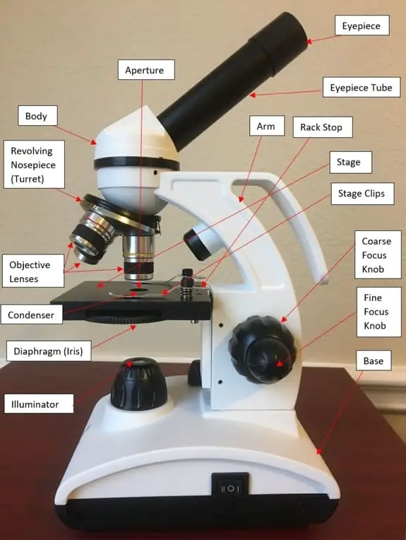
Compound Microscope Definitions for Labels. Eyepiece (ocular lens) with or without Pointer: The part that is looked through at the top of the compound microscope. Eyepieces typically have a magnification between 5x & 30x. Monocular or Binocular Head: Structural support that holds & connects the eyepieces to the objective lenses.
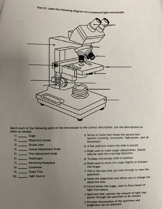
Solved Part Iii Label The Following Diagram Of A Compound Chegg Com
Microscope Description A microscope is a laboratory instrument used to examine objects that are too small to be seen by the naked eye. In other words, it enlarges images of small objects. Invented by a Dutch spectacle maker in the late 16th century, light microscopes use lenses and light to magnify images. Generally a microscope ... Read more 22 Parts Of a Microscope With Their Function And ...
Simple Light Microscope Diagram Clip Art Library
Plant Cells Under Microscope Green Pattern Plant Cell Plant Pattern Histolab4a Htm Cell Biology Teaching Biology Biology Lessons Pin On My Universe Microscope Images Of Plant Seeds And Pollens Rapeseed Brassica Napus Pollen Grain Microscopic Images Electron Microscope Images Science Photos Plant Cell Electron Microscope Worksheet Plant Cell Cell Diagram Plant Cell Diagram Jamesalliban Biology

Microscope Labeling
admin January 4, 2021. Some of the worksheets below are Parts and Function of a Microscope Worksheets with colorful charts and diagrams to help students familiarize with the parts of the microscope along with several important questions and activities with answers. Basic Instructions. Once you find your worksheet (s), you can either click on ...
Microscope Labeling Activity Smart Board Activity By Mrs Flynn Science
Click to Download : Label the Parts of the Microscope with answers (A4) PDF print version. For a thorough review of each microscope part continue reading…. A basic microscope has a single convex lens such as those found in a magnifying glass, which you can use to visualize the finest prints.

Microscope Diagram Labeled Unlabeled And Blank Parts Of A Microscope
Nov 14, 2018 - A collection of microscope diagrams and worksheets for science class. ... FREE Parts of a Microscope Label Worksheet. More information.
16 Parts Of A Compound Microscope Diagrams And Video Microscope Clarity
1. Eyepiece/Ocular Lens – The lens into which the user looks to see the specimen. ... 3. Arm – A supporting piece of the optical microscope mounted upon the base.

Compound Microscope Parts Functions And Labeled Diagram New York Microscope Company
Jejunum histology slide labeled diagram. Now, it is better to review all the histological features from the jejunum slide. I will try to show you all the features from the jejunum histology slide with the labeled diagram. Here, I tried to show you all the histological features from the four different layers of the jejunum wall.

Diagram Of A Compound Microscope
Labeled diagram of a compound microscope. The ocular lens, the objective lens, the iris diaphragm — all these pieces work together to magnify the they may also be printed as teacher resources. A standard microscope has three four or five objective lenses that each microscope layout both blank and the version with answers are available as pdf ...

Compound Microscope Definition Labeled Diagram Parts Uses
Jan 13, 2016 - Free worksheets for labeling parts of the microscope including a ... Microscope Diagram (Grade 8) - Free Printable Tests and Worksheets.

Compound Monocular Microscope Clear Imaging Biological Microscope Price China Monocular Microscope Binocular Microscope Made In China Com
Image : Labeled Diagram of compound microscope parts. See: Labeled Diagram showing differences between compound and simple microscope parts Structural Components. The three structural components include. 1. Head. This is the upper part of the microscope that houses the optical parts. 2. Arm This part connects the head with the base and provides ...

Compound Microscope Diagram Structure And Principle
Figure: Diagram of parts of a microscope. There are three structural parts of the microscope i.e. head, base, and arm. Head - This is also known as the body, it carries the optical parts in the upper part of the microscope. Base - It acts as microscopes support. It also carries microscopic illuminators.

Microscope With Labels Svg Vector Microscope With Labels Clip Art Svg Clipart
To look at a cell close up we need a microscope. Dec 07, 2020. Plant Cell Labeled Under Microscope : Plant cells have cell walls, one large vacuole per cell, and chloroplasts, while animal cells will have a cell membrane only. Original Resolution: 350x275 px. 21 Plant Cell Facts For Kids Learn About Plant Cells - Contact microscope world with ...

Solved Tration Questions 10 Points Label The Diagram Of A Chegg Com
Image Result For Diagram Of Plant And Animal Cell Under Electron Microscope Celula Animal Ciencias Verdades Absolutas. 560 X 364 Pixel Electron Microscope Image Animal Cell And Organelles Labeled Animal Cell Plasma Membrane Organelles. Royalty Free Stock Photos A Typical Cell Labeled Cell Diagram Animal Cell Cells Worksheet.

Compound Microscope Parts Labeled Diagram And Their Functions Rs Science
Compound Light Microscope Diagram. A Study Of The Microscope And Its Functions With A Labeled Diagram. Free Microscope Drawing Download Free Clip Art Free Clip Art On. Light Microscope Sketch At Paintingvalley Com Explore Collection. Compound Microscope Drawing Clipart Library Clip Art Library.

Carl Zeiss Microscopy Optical Microscope Worksheet Diagram Microscope Angle Technic Png Pngegg
40x 400x Compound Monocularbiological Microscope45 Degree Angled Headelectric Lightedbeginner Slides Plant Cell Things Under A Microscope Plant Cell Picture. Plasmolysis Of Elodea Biology Experiments Apologia Biology Teaching Science. Elodea Leaf Cell Under Microscope Plant Cell Cells Worksheet Lab Activities.

Schematic Diagram Showing Principle Of Compound Light Microscope Download Scientific Diagram
11. Obtain a prepared microscope slide of a nerve. Locate the cross section of the nerve, and note the many round nerve fibers inside. Nerve fiber is a. Figure 25.1 Label this diagram of a motor neuron. Figure 25.1 Label this diagram of a motor neuron. Figure 25.2 Label the features of the myelinated nerve fiber.

Compound Microscope Diagram Parts Labelled Principle And Uses Microscope Wiki

Label Microscope Diagram Enchantedlearning Com
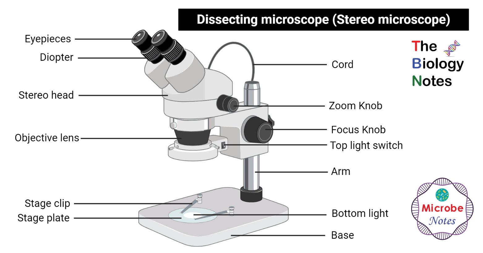
Dissecting Microscope Stereo Or Stereoscopic Microscope

Microscope Parts And Functions

White And Black Microscope Optical Microscope Light Scanning Electron Microscope Diagram Microscope Angle Technic Laboratory Png Pngwing
Premium Vector Microscope With Plant Cell Diagram

Microscope Labeled Diagram

Labelling A Microscope Diagram Quizlet

Using The Microscope Docsbay

Microscope Types Parts History Diagram Facts Britannica

Simple Microscope Diagram Parts Labelled Principle Formula And Uses Microscope Wiki
Microscope Using A Microscope I Have Developed My
1

Microscope Diagram And Quiz Science Diagrams Science Printables Science Teaching Resources
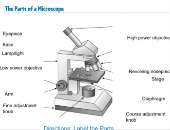
Label A Microscope Teaching Resources
0 Response to "37 diagram of a microscope with labels"
Post a Comment