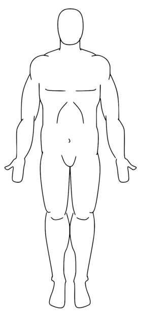
37 anatomical position diagram blank
Anatomical position, h Posterior, i.Anterior, j. Dorsal, k.Ventral. ANATOMICAL PLANES OF THE BODY Many specimens in anatomyare sectioned so that the interior of the organ or region can be examined. It is important that the direction of the cut is known so thatthe proper Sep 30, 2019 · Anatomical position is the description of any region or part of the body in a specific stance. In the anatomical position, the body is upright, directly facing the observer, feet flat and directed forward. The upper limbs are at the body’s sides with the palms facing forward. Like so: Image captured in AR using Human Anatomy Atlas.
Ventral/Dorsal–Equivalent to belly-side and back-side of a body in anatomical position. For a human in anatomical position, this pair of terms is equivalent to anterior and posterior. However, for four-legged animals in what is considered their anatomical position, …
Anatomical position diagram blank
May 12, 2019 · Anatomical Position. The kidneys lie retroperitoneally (behind the peritoneum) in the abdomen, either side of the vertebral column.. They typically extend from T12 to L3, although the right kidney is often situated slightly lower due to the presence of the liver.Each kidney is approximately three vertebrae in length. The adrenal glands sit immediately superior to the kidneys within a separate ... Diagram the forces acting on the combination of gymnast Jun 19, 2018 · Drag the pink labels to the pink targets to indicate the sex dictated by the genotype in each box. Open an wiring diagram example or a blank drawing page. Drag the labels onto the diagram to identify the various chromosome structures. Labels can be used once or more than once. Unlabelled Diagrams for Human Anatomy BIO 26-211 Fig. 19.20 Fig. 25.2a Fig. 26.12a Fig. 27.3 Fig. 28.2 Fig. 28.3a Fig. 28.11 Fig. 28.15a Unlabelled Diagrams for Human ...
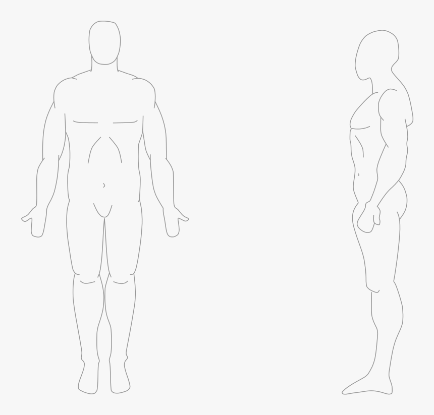
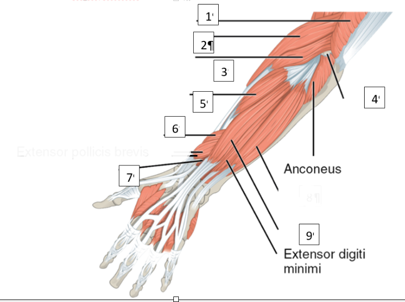
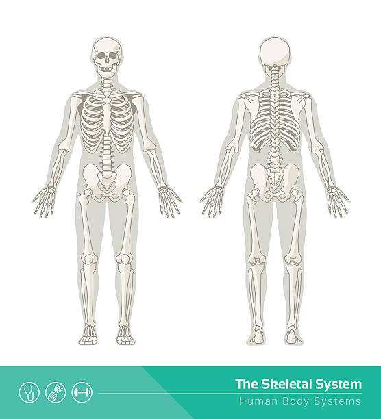
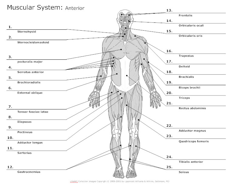
Anatomical position diagram blank. Anatomy worksheets are an illustration of a certain part or system of the body, with 'fill in the blank' spaces pointing to different sections of the illustration. Anatomy charts can be specific to one part of the body, such as a knee joint, or cover a combination of body parts: the skeletal system, for example. 9+ Free Body Diagram – Free Printable Download. Studying the structure of a human body without visual aid is quite complicated. ... The students of today need understanding, so, you cannot teach a topic like this without a body diagram template. The body diagram template you download depends on the part of the body you want to talk about ... The compartments of the distal upper extremity include the superficial anterior, deep anterior, and posterior regions. The muscles of the anterior chamber all act to flex the wrist (and usually include the term "flexor" in their name) while the muscles of the posterior chamber act to extend the wrist (and thus include the term "extensor") in their name. When filling out a human body outline worksheet, it is vital that you use some sort of a reference like a text book to ensure that the information you are putting on your outline is correct.There are a few different kinds of outlines you can choose from. An example of one would be a human body outline template with organs, this is perfect for someone that needs to know where and what all the ...
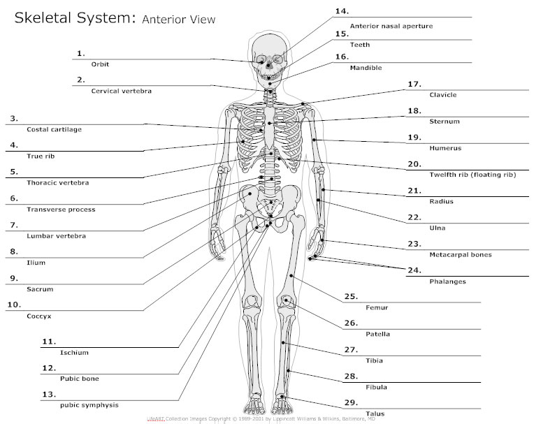
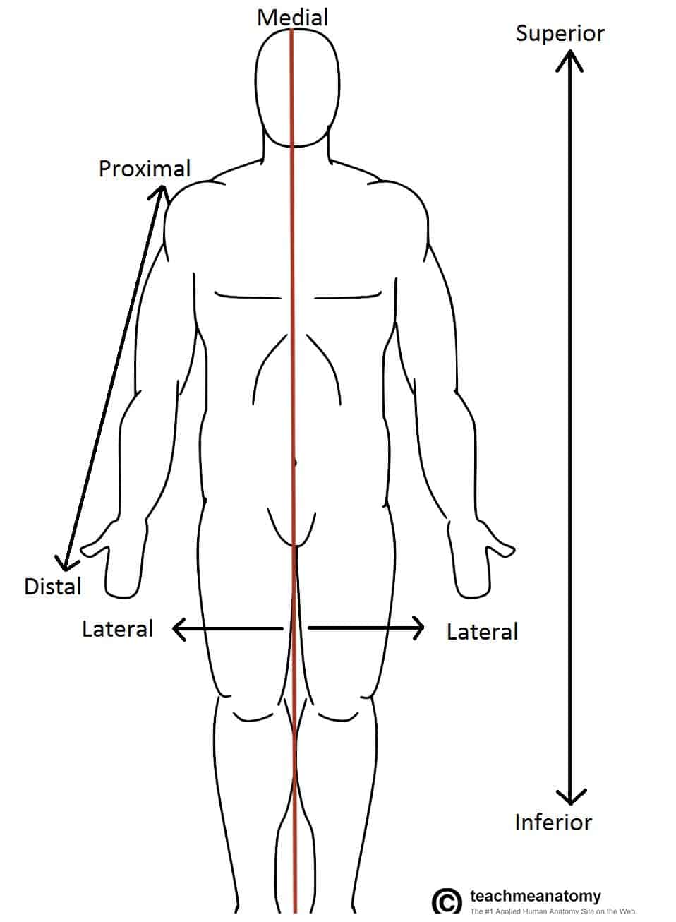
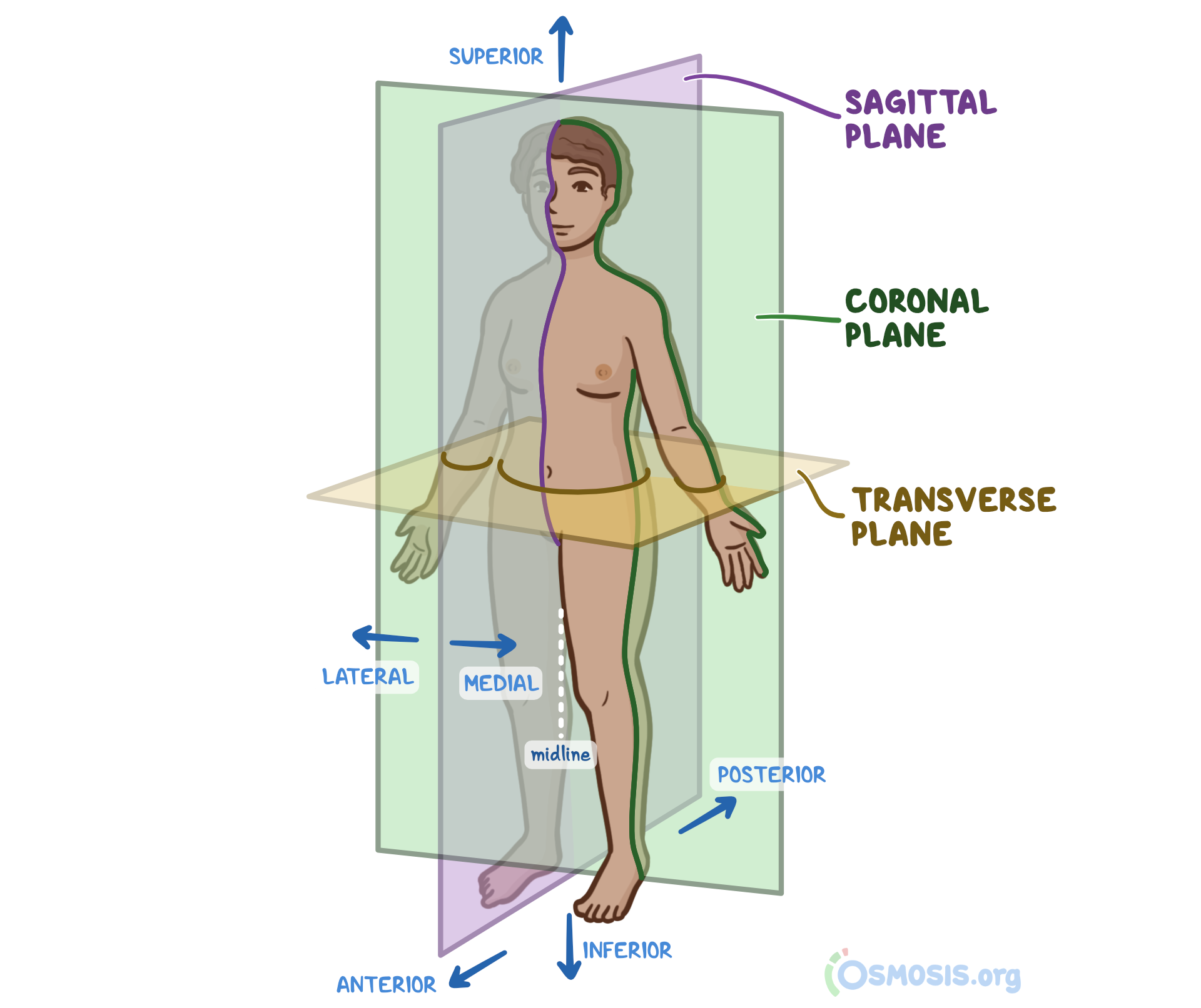
Insert your answer in the blank IR.low the ... 10 color the coding Circles and the corresponding structures in the diagram. Then. label the angles indicated by leader lines. 85 Acromion Lateral border ... Medial hone of the forearm in anatomical position 14. 15] Rounded … BIO 113 LAB 1. Anatomical Terminology, Positions, Planes, and Sections and more . Objectives • Describe the anatomical position verbally or by demonstrating it • Demonstrate ability to use anatomical terms describing body landmarks, directions, planes, and surfaces. • Name the body cavities, and indicate important organs in each cavity. Anatomical Planes The anatomical position is further standardized by dividing the body into three anatomical planes. A plane is an imaginary flat surface passing through the body or organ which divides the structure. 1) Frontal (Coronal) Plane: is vertical and extends from one side of the body to the other. It divides the body into front and ... The Anatomical Position. First, let’s talk about the anatomical position. The anatomical position is a standing position, with the head facing forward and the arms to the side. The palms are facing forward with the fingers extended, and the thumbs are pointing away from the body. The feet are spaced slightly apart with the toes pointing forward.
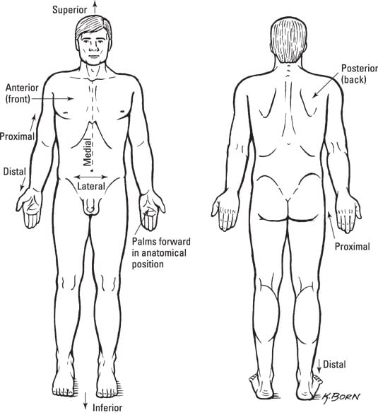
Sep 15, 2021 · Explore more about anatomical directional terminology and the four terms that reference the position from the midline. Updated: 09/15/2021 Create an account May 11, 2020 · Diagram of a ring clasp. Diagrams of gingivally approaching clasps Considerations when choosing a clasp - Summary. Position of undercut – dependent on your final survey line! Remember lower molars may be more undercut on the lingual side due to their inclination- this would be the retentive surface. Health of PDL – consider recession Anatomical Position. To further increase precision, anatomists standardize the way in which they view the body. Just as maps are normally oriented with north at the top, the standard body “map,” or anatomical position, is that of the body standing upright, with the feet at shoulder width and parallel, toes forward. Anatomical Position. To further increase precision, anatomists standardize the way in which they view the body. Just as maps are normally oriented with north at the top, the standard body “map,” or anatomical position, is that of the body standing upright, with the …
Anatomical positions are important because they give us a frame of reference for describing the body. Similar to a compass, they give us a universal way to describe the position of an organism. The concept of anatomical position is particularly important in medicine , as mistakes can occur if medical professionals do not have a shared point of ...
Dec 23, 2020 · The brachial plexus is a network of nerve fibres that supplies the skin and musculature of the upper limb. It begins in the root of the neck, passes through the axilla, and runs through the entire upper extremity. The plexus is formed by the anterior rami (divisions) of cervical spinal nerves C5, C6, C7 and C8, and the first thoracic spinal nerve, T1.
Unlabelled Diagrams for Human Anatomy BIO 26-211 Fig. 19.20 Fig. 25.2a Fig. 26.12a Fig. 27.3 Fig. 28.2 Fig. 28.3a Fig. 28.11 Fig. 28.15a Unlabelled Diagrams for Human ...

Diagram the forces acting on the combination of gymnast Jun 19, 2018 · Drag the pink labels to the pink targets to indicate the sex dictated by the genotype in each box. Open an wiring diagram example or a blank drawing page. Drag the labels onto the diagram to identify the various chromosome structures. Labels can be used once or more than once.
May 12, 2019 · Anatomical Position. The kidneys lie retroperitoneally (behind the peritoneum) in the abdomen, either side of the vertebral column.. They typically extend from T12 to L3, although the right kidney is often situated slightly lower due to the presence of the liver.Each kidney is approximately three vertebrae in length. The adrenal glands sit immediately superior to the kidneys within a separate ...


:max_bytes(150000):strip_icc()/anatomical-directional-terms-and-body-planes-373204-01-28f62bf8f2214f71b9d38dfa85beccb5.png)

0 Response to "37 anatomical position diagram blank"
Post a Comment